A child tugging at their ear, a patient with a sudden nosebleed – understanding common UKMLA ENT presentations is a cornerstone of general practice and a key area for your UK Medical Licensing Assessment (UKMLA). Are you prepared to confidently assess and manage them? ENT conditions are highly prevalent in primary care and emergency settings, forming a significant part of what a Foundation Year doctor will encounter. The General Medical Council’s (GMC) MLA content map, which outlines the core knowledge, skills, and behaviours needed for UK practice, explicitly includes “Ear, nose and throat” as an area of clinical practice. This assessment focuses on common and acute presentations, ensuring candidates are ready for safe practice. Indeed, mastering these scenarios directly aligns with the three overarching themes of the UKMLA: Readiness for safe practice, Managing uncertainty, and Delivering person-centred care.
The breadth of the UKMLA syllabus can feel daunting, and the pressure to perform is understandable. This post aims to be a focused, high-yield resource, streamlining your revision of key UKMLA ENT presentations by providing a practical, guideline-based approach to common conditions. As the GMC notes, “The MLA content map – which includes an ‘A-Z of presentations’ that may be assessed in the exam – is useful for identifying topics which you may need to revise”. Understanding these presentations is therefore directly relevant to exam success. It’s important to recognize that the UKMLA isn’t merely testing factual recall of ENT conditions; it’s assessing a candidate’s ability to integrate this knowledge with clinical reasoning and professional behaviours, as outlined in the GMC’s “Outcomes for Graduates”. The content map specifically concentrates on “professional skills, knowledge and behaviours that are essential for safe practice”. Therefore, excelling in the ENT component of the UKMLA means demonstrating the application of knowledge in a safe, professional, and patient-centred manner. This guide will cover key conditions like otitis media, tonsillitis, epistaxis, and sinusitis, equipping you with the essentials for both the exam and your future practice.
Key Takeaways
- Mastering common UKMLA ENT presentations like otitis media, tonsillitis, epistaxis, and sinusitis is crucial for exam success and safe Foundation Year practice.
- National Institute for Health and Care Excellence (NICE) guidelines are central to managing ENT conditions in the UK; understanding their recommendations on diagnosis, antibiotic stewardship, and referral is key.
- Scoring systems like FeverPAIN (for tonsillitis) aid in clinical decision-making and support the appropriate use of antibiotics.
- Recognising red flags in ENT presentations is vital for patient safety and ensuring timely specialist referral.
- The UKMLA emphasizes patient-centred care, safe practice, and managing uncertainty – principles that are directly applicable to all ENT encounters.
- Effective communication and adherence to antimicrobial stewardship principles are integral to managing ENT conditions as per UK standards.
- Understanding the rationale behind guideline recommendations, such as watchful waiting or delayed prescribing, demonstrates a deeper grasp of evidence-based medicine.
Acute Otitis Media (AOM) & Otitis Media with Effusion (OME): UKMLA Clinical Scenarios
Acute Otitis Media (AOM) is defined as an acute inflammation or infection of the middle ear, while Otitis Media with Effusion (OME), commonly known as glue ear, refers to a collection of sterile fluid in the middle ear space. Both conditions are highly prevalent, particularly in children, making them frequent encounters in primary care and important topics for UKMLA ENT presentations. A key challenge for candidates is differentiating between AOM and OME, as their management pathways differ significantly.
Typical Presentations
The clinical presentation of AOM typically includes otalgia (earache), fever, irritability, and hearing loss. If the tympanic membrane perforates, ear discharge may be present. Young children with AOM may exhibit non-specific symptoms such as ear rubbing, reduced feeding, or restlessness. In contrast, OME is often asymptomatic or may present more insidiously with hearing impairment (conductive in nature), which can sometimes lead to speech delay in children, or a sensation of fullness in the ear. OME frequently follows an upper respiratory tract infection (URTI) or an episode of AOM. The common presentations of otalgia and deafness underscore the importance of distinguishing these conditions.
Key Diagnostic Features & Otoscopic Findings
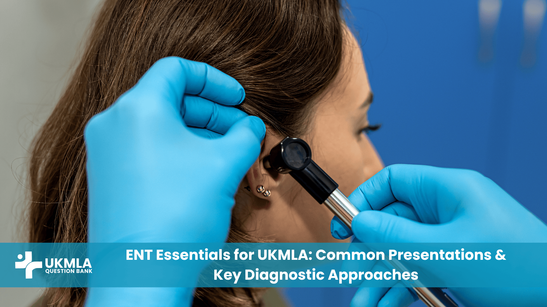
Otoscopy is a fundamental skill, noted as a practical procedure in the UKMLA syllabus , and is crucial for diagnosis.
- In AOM, otoscopic examination typically reveals a bulging, erythematous tympanic membrane (TM) with loss of the normal light reflex. A purulent effusion may be visible behind the TM, or there might be evidence of perforation with active discharge.
- For OME, the TM often appears retracted or in a neutral position and may be opacified or dull. Air-fluid levels or bubbles might be visible behind the TM, and its mobility may be impaired on pneumatic otoscopy (if available).
NICE Guideline Summary (Diagnosis & Management)
Adherence to NICE guidelines is paramount in UK practice.
AOM (NICE Guideline NG91): Most cases of AOM are viral and self-limiting, typically resolving within about three days without specific antibiotic treatment. Management should focus on providing adequate analgesia, such as paracetamol or ibuprofen. Antibiotics provide limited benefit for most children and are not routinely recommended. NICE NG91 emphasizes an antimicrobial prescribing strategy aimed at limiting antibiotic use and reducing antimicrobial resistance. A delayed or back-up antibiotic prescription strategy is often appropriate, with clear advice given to parents or patients on when to use it. Immediate antibiotics are generally reserved for specific situations, such as when the patient is systemically unwell, at high risk of complications (e.g., due to significant co-morbidities), in children under two years with bilateral AOM, or if there is otorrhoea (ear discharge) due to TM perforation. If an antibiotic is indicated, phenoxymethylpenicillin is the first-line choice, with clarithromycin or erythromycin (particularly in pregnancy) as alternatives for those with penicillin allergy. For further details, refer to the NICE NG91 guideline on acute otitis media.
OME : For OME, a period of “watchful waiting” for up to three months is common, as many cases resolve spontaneously. If symptoms, particularly hearing loss, persist beyond three months, or if there are concerns about the child’s development (such as speech delay or impact on school attainment), a hearing assessment is recommended. Autoinflation techniques, using devices like a nasal balloon, may be considered for some children as a non-surgical management option. Importantly, antibiotics, oral or nasal corticosteroids, antihistamines, and decongestants are NOT routinely recommended for the management of OME, as evidence suggests they offer clinically insignificant improvement. Referral to an ENT specialist for consideration of grommet insertion (tympanostomy tubes) may be appropriate if there is persistent bilateral OME associated with significant hearing loss or developmental impact.
The careful consideration of whether to prescribe antibiotics in AOM, often opting for delayed prescribing, or the decision for watchful waiting in OME, reflects an ability to handle the inherent uncertainties in clinical presentations. This approach aligns with the UKMLA’s theme of “managing uncertainty” , where practitioners must make sound clinical judgments based on evolving symptoms and guideline adherence, rather than resorting to immediate, potentially unnecessary interventions. This judicious approach, particularly the strong emphasis on antimicrobial stewardship in AOM and the avoidance of ineffective treatments in OME , also demonstrates an understanding of broader NHS principles regarding resource optimization and combating antimicrobial resistance. Such awareness is integral to being “ready for safe practice” within the UK healthcare system.
Red Flags & Referral Criteria (AOM & OME)
Recognising red flags is crucial for patient safety:
AOM: Signs suggestive of mastoiditis (such as post-auricular swelling, erythema, tenderness, or outward and downward displacement of the pinna), facial nerve palsy, signs of meningitis (e.g., neck stiffness, photophobia, altered mental state), severe systemic illness, or persistent symptoms despite antibiotic treatment warrant urgent referral.
OME: Persistent hearing loss that is impacting a child’s speech, language, or educational development, or suspicion of associated conditions (e.g., children with Down’s syndrome or cleft palate, who are at increased risk of prolonged glue ear and may benefit from earlier referral) are indications for specialist assessment.
Table 1: Differentiating AOM and OME: Symptoms, Signs, and Initial Management (NICE-aligned)
This table provides a clear, side-by-side comparison essential for rapid differentiation in exam questions and clinical practice.
| Feature | Acute Otitis Media (AOM) | Otitis Media with Effusion (OME) | Initial Management Principles (NICE-aligned) |
|---|---|---|---|
| Onset | Acute, often rapid | Gradual or asymptomatic; may follow AOM/URTI | AOM: Assess severity. OME: Often watchful waiting. |
| Otalgia (Earache) | Prominent, often severe | Mild, absent, or feeling of fullness | AOM: Prioritise analgesia (paracetamol/ibuprofen). OME: Analgesia if discomfort. |
| Fever | Common, can be high | Usually absent or low-grade | AOM: Antipyretics for fever. |
| Hearing Loss | Often present, conductive | Key feature, conductive; may fluctuate | AOM: Usually transient. OME: Monitor impact, formal hearing test if persists >3 months or concerns. |
| TM Appearance | Bulging, erythematous, loss of light reflex; possible pus/perforation | Retracted/neutral, dull/opacified, air-fluid levels/bubbles, impaired mobility | AOM: Key diagnostic sign. OME: Key diagnostic sign. |
| Systemic Symptoms | Possible (irritability, lethargy, poor feeding in infants) | Usually absent | AOM: Assess for systemic illness (immediate antibiotics if present). |
| Typical Patient | Often young children | Common in children, can affect adults | AOM: Follow NICE NG91. OME: Watchful waiting, consider autoinflation; avoid routine antibiotics/steroids. |
| Management Focus | Symptomatic relief, judicious antibiotic use, monitor for complications | Monitoring hearing and development, consider grommets if persistent and impactful | AOM: Antimicrobial stewardship is key.[9] OME: Active monitoring, referral for persistent issues affecting development. |
Sore Throat & Tonsillitis: Applying NICE Guidelines for UKMLA Success
Sore throat is an exceedingly common presentation in primary care, and tonsillitis, characterized by inflammation of the tonsils, is a frequent cause of severe sore throat. The vast majority of these cases are viral in origin and self-limiting. For the UKMLA candidate tackling UKMLA ENT presentations like tonsillitis, the primary challenge lies in correctly identifying the small proportion of patients who may benefit from antibiotic therapy, adhering to principles of antimicrobial stewardship, and promptly recognizing potential complications that necessitate further action.
Common Presentations & Differentiating Viral vs. Bacterial
Patients with tonsillitis typically present with a sore throat, odynophagia (pain on swallowing), fever, visibly inflamed tonsils which may have exudate, and tender cervical lymphadenopathy. Differentiating between viral and bacterial causes can be challenging but is important for guiding management.
- Features suggestive of a viral etiology often include coryzal symptoms (runny nose, sneezing), cough, hoarseness, and sometimes diarrhoea; the illness may be generally milder.
- Conversely, features more indicative of bacterial tonsillitis, particularly Group A Streptococcus (GAS), include a more sudden onset, marked fever, visible tonsillar exudate, tender anterior cervical lymph nodes, and a notable absence of cough. These features form the basis of the Centor criteria.
Diagnostic Scoring Systems: FeverPAIN and Centor Criteria
To aid in predicting the likelihood of GAS infection and to guide antibiotic prescribing decisions, clinical scoring systems are employed. NICE recommends the use of the FeverPAIN score.
The Centor Criteria include :
- Tonsillar exudate
- Tender anterior cervical lymphadenopathy
- History of fever (over 38°C)
- Absence of cough
A score of 3-4 suggests a higher likelihood of GAS.
The FeverPAIN Criteria, which NICE advocates, are :
- Fever (in the previous 24 hours)
- Purulence (pharyngeal/tonsillar exudate)
- Attend rapidly (patient presents within 3 days of symptom onset)
- Severely Inflamed tonsils
- No cough or coryza
Each criterion scores one point. The FeverPAIN scoring system is designed to “help identify those who are most likely to benefit from antibiotics”.
NICE Recommendations for Management
NICE guidance emphasizes symptomatic relief and judicious antibiotic use.
- Symptomatic relief: Analgesia with paracetamol or ibuprofen is valuable for managing pain and fever. Patients should also be advised on self-care measures such as rest and adequate fluid intake.
- Antibiotics: For most patients, antibiotics have a limited impact, shortening the duration of symptoms by only approximately 16 hours overall.
- Antibiotic Prescribing based on FeverPAIN score :
- Score 0-1: Do not offer an antibiotic.
- Score 2-3: Consider no antibiotic or a delayed/back-up antibiotic prescription. (For adults with a score of 2 or 3, no severe symptoms, and not in a vulnerable group, a delayed prescription is an option).
- Score 4-5: Consider an immediate antibiotic prescription. (For children with a score of 3 or more, prescribe antibiotics).
- Choice of Antibiotic :
- The antibiotic of choice is phenoxymethylpenicillin, typically for a 5-10 day course. While 5 days may be sufficient for symptomatic relief in some, a 10-day course is traditionally recommended for proven GAS infection to prevent complications like rheumatic fever.
- For individuals with penicillin allergy or intolerance, a five-day course of clarithromycin is the alternative first choice. Erythromycin is preferred in pregnancy if there is a penicillin allergy.
- Amoxicillin should be avoided if there is a possibility of glandular fever (infectious mononucleosis) due to the risk of inducing a characteristic maculopapular rash.
NICE also recommends that “a back-up antibiotic prescription may be considered…[with advice on when to use it]”. This practical advice, including when to start the antibiotic if symptoms don’t improve or worsen, is crucial for UKMLA scenarios and patient communication.
Highlighted Key Point 1: The crucial role of FeverPAIN/Centor scores in guiding antibiotic decisions for tonsillitis, aligning with antimicrobial stewardship principles. For UKMLA candidates, correctly applying these scores demonstrates an understanding of evidence-based medicine and the judicious use of antibiotics, key tenets of safe UK practice.
The application of scoring systems like FeverPAIN moves beyond simple protocol adherence. It facilitates a more tailored approach to antibiotic decisions based on an individual’s risk profile. This nuanced decision-making, which avoids unnecessary medication and potential side effects for those unlikely to benefit, is a clear example of “delivering person-centred care,” a core theme of the UKMLA. This patient-centric approach ensures that treatment is appropriate for the individual, rather than a one-size-fits-all strategy. Furthermore, the evolution of guidelines towards more restrictive antibiotic use for tonsillitis reflects how healthcare systems adapt to emerging evidence on antimicrobial resistance and treatment efficacy. UKMLA candidates are expected to practice within this dynamic, evidence-based framework, demonstrating an understanding that medical practice changes as new evidence becomes available.
Red Flags & Complications
It is vital to recognise warning signs of complications:
- Peritonsillar abscess (quinsy): Characterised by severe unilateral sore throat, trismus (difficulty opening the mouth), dysphagia, a muffled “hot potato” voice, and often deviation of the uvula away from the affected side. This is a clinical emergency requiring urgent referral for assessment and potential drainage.
- Airway compromise: Stridor (a high-pitched breathing sound), difficulty breathing, or drooling are ominous signs requiring immediate emergency management.
- Systemic sepsis: Signs such as high fever, tachycardia, hypotension, or confusion.
- Glandular fever (infectious mononucleosis): Should be considered, especially in adolescents and young adults, if there is a prolonged sore throat accompanied by significant fatigue, widespread lymphadenopathy, and fever. A Paul-Bunnell test or monospot test can aid diagnosis.
- Scarlet fever: If tonsillitis is caused by GAS and is accompanied by the characteristic sandpaper-like rash, a 10-day course of antibiotics is required to manage the scarlet fever and prevent complications.
Indications for Referral/Tonsillectomy
Referral to ENT may be necessary in certain situations:
- Emergency referral: For suspected airway obstruction, peritonsillar abscess (quinsy), or severe systemic sepsis.
- Elective referral for tonsillectomy (NICE criteria for recurrent tonsillitis ):
- Seven or more clinically significant, adequately treated sore throats in the preceding year.
- Five or more such episodes in each of the preceding two years.
- Three or more such episodes in each of the preceding three years.
- The episodes of sore throat must be disabling and prevent normal functioning.
Epistaxis (Nosebleeds): A Practical Approach for UKMLA Candidates
Epistaxis, or bleeding from the nose, is a very common complaint encountered in various healthcare settings. While most episodes are benign and self-limiting, they can be alarming for patients and, on occasion, can be severe, requiring prompt and effective management. For UKMLA candidates addressing UKMLA ENT presentations such as epistaxis, a systematic approach to assessment, knowledge of immediate management techniques, and understanding when to escalate care or refer are essential.
Common Causes and Sites
A multitude of factors can precipitate epistaxis :
- Trauma: This is the most frequent cause, with nose picking being particularly common, especially in children. Other forms of trauma include insertion of foreign bodies or excessive nose blowing, often associated with URTIs when the nasal mucosa is congested.
- Mucosal Dryness: Environmental factors such as central heating or cold, dry weather can lead to desiccation and cracking of the nasal mucosa.
- Inflammation: Conditions like URTIs, sinusitis, and allergic rhinitis can cause nasal mucosal inflammation and increase friability.
- Medications: Anticoagulants (e.g., warfarin, DOACs) and antiplatelet drugs (e.g., aspirin, clopidogrel) can predispose to or prolong bleeding.
- Bleeding Disorders: Systemic conditions affecting haemostasis, such as thrombocytopenia, von Willebrand disease, or other coagulopathies, can manifest with epistaxis.
- Hypertension: While not always a direct cause, high blood pressure can prolong or exacerbate an episode of epistaxis.
- Less Common Causes: These include vascular abnormalities like hereditary haemorrhagic telangiectasia (Osler-Rendu-Weber syndrome), nasal tumours (e.g., juvenile nasopharyngeal angiofibroma, typically seen in adolescent males), and illicit drug use (e.g., cocaine insufflation, which can cause septal damage).
The site of bleeding is a key determinant of severity and management :
- Anterior Epistaxis: Accounts for approximately 90% of cases. Bleeding typically originates from Kiesselbach’s plexus (also known as Little’s area), a rich anastomosis of blood vessels on the anterior nasal septum. Anterior bleeds are generally easier to control.
- Posterior Epistaxis: Less common (around 10%) but often more profuse and challenging to manage. Bleeding usually arises from branches of the sphenopalatine artery. Blood may be seen running down the posterior pharynx, and there is a higher risk of aspiration and airway compromise.
Systematic Assessment
A thorough assessment is crucial:
- History: Enquire about the onset, duration, and frequency of bleeding, estimate the amount of blood loss, determine if it’s unilateral or bilateral, and identify any provoking factors. A detailed medical history, including any known bleeding disorders, hypertension, current medications (especially anticoagulants/antiplatelets), and family history of bleeding problems, is vital.
- Examination:
- In cases of severe bleeding, an ABCDE (Airway, Breathing, Circulation, Disability, Exposure) approach should be adopted first.
- Monitor vital signs, including pulse and blood pressure.
- Once initial control measures are in place, attempt to identify the bleeding site (anterior or posterior) using a good light source (e.g., headlamp) and a nasal speculum.
Immediate Management (First Aid & Clinical Setting)
Effective immediate management can often resolve the bleeding:
First Aid Measures :
- The patient should sit upright and lean slightly forward. This position helps prevent blood from being swallowed (which can cause nausea and vomiting) and reduces venous pressure in the nasal vessels.
- Firmly pinch the soft, fleshy part of the nose (anterior nares) continuously for 10-20 minutes.
- Encourage the patient to breathe through their mouth.
- Any blood in the mouth should be spat out.
- An ice pack applied to the bridge of the nose or the nape of the neck may help promote vasoconstriction.
Clinical Setting (if first aid fails):
- Ensure all necessary equipment is readily available: good lighting (headlight), suction, nasal speculum, topical anaesthetic and vasoconstrictor spray (e.g., cophenylcaine, which contains lidocaine and phenylephrine, though age restrictions apply ), silver nitrate sticks for cautery, and nasal packing materials.
- After applying topical spray (if used), wait a few minutes for it to take effect, then gently attempt to locate the bleeding point.
“The MLA content map concentrates on the professional skills, knowledge and behaviours that are essential for safe practice – for example, effective communication, recognising risk and the diagnosis and management of common or acute presentations and conditions.” (Adapted from ).
Managing epistaxis effectively, which includes providing clear instructions to the patient or carers (effective communication) and identifying when simple first-aid measures are insufficient or when the bleeding is severe (recognising risk), directly aligns with these core UKMLA competencies.
NICE CKS Guidance on Cautery & Packing
If bleeding persists despite first-aid measures, further interventions may be required, guided by principles outlined in resources like NICE Clinical Knowledge Summaries (CKS).
- Nasal Cautery (Silver Nitrate):
- This is suitable for visible anterior bleeding points once active bleeding has been controlled or significantly reduced.
- Apply the tip of a silver nitrate stick directly to the bleeding vessel for approximately 5-10 seconds. Work from the edge of the bleeding area inwards.
- A crucial safety point is to avoid cauterizing both sides of the nasal septum during the same session, as this carries a risk of septal perforation.
- Following cautery, particularly for recurrent bleeds, application of a topical antiseptic cream (e.g., Naseptin®, containing chlorhexidine and neomycin) twice daily for a week or two can help prevent infection and promote healing.
- Anterior Nasal Packing:
- Indicated if bleeding continues despite sustained pressure and cautery is not possible, not effective, or the bleeding point cannot be identified.
- Various materials can be used, including prefabricated nasal tampons (e.g., Merocel® , Rapid Rhino®) which expand on contact with moisture, or ribbon gauze impregnated with petroleum jelly or bismuth iodoform paraffin paste (BIPP).
- The pack should be inserted along the floor of the nasal cavity. Ensure it is firm enough to apply pressure but avoid over-packing, which can cause pain and pressure necrosis.
- If an anterior pack successfully controls the bleeding, the patient may be discharged with instructions for follow-up, typically within 24-48 hours, for pack removal by a healthcare professional.
- Prophylactic oral antibiotics are sometimes considered if a nasal pack is to remain in situ for more than 24 hours to reduce the risk of local infection or toxic shock syndrome, though practice varies.
- Posterior Packing: This is a more complex procedure, usually reserved for suspected posterior bleeds that are not controlled by anterior packing. It typically requires specialist ENT input and may involve inflatable balloon catheters or formal gauze packs. Patients requiring posterior packing usually need hospital admission.
The decision to proceed with interventions like cautery or packing involves not only knowledge of the techniques but also an appreciation of the practical skills required and the potential risks, such as septal perforation with bilateral cautery. This underscores that the UKMLA assesses not just factual recall but also procedural understanding and a strong emphasis on safety consciousness, reflecting the practical skills component of the syllabus. When managing epistaxis in elderly patients or those on anticoagulants , a holistic assessment is critical. It’s not merely about stopping the nosebleed but involves considering the patient’s wider medical context, comorbidities, and medications. This broader view, which might involve liaising with other specialists or carefully weighing the risks of reversing anticoagulation, is a hallmark of “delivering person-centred care” and is an implicit expectation for UKMLA candidates.
Table 2: Step-by-Step Management of Acute Epistaxis in a Primary Care/Emergency Setting (NICE CKS based)
This table provides a structured, algorithmic approach to a common acute presentation.
| Step | Action | Key Considerations/Timings |
|---|---|---|
| 1. Initial Assessment | ABCDE if severe. History (frequency, amount, laterality, trauma, meds). Vital signs. | Prioritise life-threatening bleeds. Identify risk factors (anticoagulants, bleeding disorders). |
| 2. First Aid Measures | Patient sits up, leans forward. Pinch soft part of nose firmly for 10-20 mins. Breathe through mouth. Spit out blood. Ice pack to nasal bridge/neck. | Ensure correct technique for nasal pressure. Continuous pressure is key. |
| 3. Reassessment & Preparation | If bleeding continues after 10-20 mins of pressure: prepare equipment (light, speculum, suction, topical vasoconstrictor/anaesthetic, cautery, packing materials). | Have all necessary equipment ready before proceeding. |
| 4. Localising Bleeding Point | Apply topical vasoconstrictor/anaesthetic if available. Gently inspect anterior nasal cavity with good light and speculum to identify bleeding site (Little’s area). | Look for active bleeding, prominent vessels, or clots. |
| 5. Anterior Bleeding Point Management (Cautery) | If a clear anterior bleeding point is seen and bleeding is not too profuse, attempt chemical cautery with silver nitrate. | Apply for 5-10 seconds. Avoid bilateral septal cautery in same session. Consider Naseptin® cream post-cautery. |
| 6. Anterior Nasal Packing | If bleeding persists despite pressure/cautery, or if cautery is not feasible, proceed with anterior nasal packing (e.g., Merocel®, ribbon gauze). | Pack firmly but carefully. Ensure pack is retrievable. Arrange follow-up for pack removal (24-48h). Consider prophylactic antibiotics if pack in situ >24h. |
| 7. When to Refer Urgently | Persistent bleeding despite adequate anterior packing; suspected posterior bleed; haemodynamic instability; recurrent severe bleeds; suspicion of tumour. | Low threshold for referral if bleeding uncontrolled, posterior source suspected, or patient unstable. Patients on anticoagulants with uncontrolled bleeding need urgent review. |
Red Flags & Referral Criteria
Prompt referral to an ENT specialist or emergency department is indicated in several situations:
- Bleeding that persists despite adequate first-aid measures and attempts at anterior cautery or packing.
- Suspected posterior bleed (e.g., blood consistently seen in the oropharynx despite anterior control, or bleeding from both nostrils that is difficult to manage).
- Haemodynamic instability (signs of shock such as tachycardia, hypotension, pallor, confusion).
- Recurrent, severe, or unexplained epistaxis, which may necessitate investigation for underlying causes like bleeding disorders or nasal pathology.
- Patients on anticoagulant or antiplatelet therapy whose bleeding cannot be controlled with standard measures.
- Suspicion of underlying malignancy (e.g., unilateral nasal obstruction, facial pain or swelling, persistent blood-stained discharge, or a visible nasal mass).
Acute Sinusitis: Diagnosis, Management, and UKMLA Pearls
Acute rhinosinusitis, commonly referred to as acute sinusitis, is an inflammation of the mucosal lining of the nose and paranasal sinuses. It is a very common condition, typically following a viral upper respiratory tract infection (URTI). While most cases are self-limiting and resolve without specific treatment, for the UKMLA candidate reviewing UKMLA ENT presentations like sinusitis, the key learning points revolve around accurate clinical diagnosis, differentiating viral from bacterial causes, providing appropriate symptomatic management, ensuring the judicious use of antibiotics according to guidelines, and recognizing potential complications that warrant urgent attention.
Recognising Typical Symptoms
The hallmark symptoms of acute sinusitis include :
- Nasal blockage or congestion: A feeling of stuffiness or obstruction in the nasal passages.
- Nasal discharge: This can be anterior (running from the front of the nose) or posterior (post-nasal drip). The discharge may initially be clear but can become thicker and purulent (yellow or green).
- Facial pain or pressure: This is often described as a dull ache or pressure sensation over the affected sinuses (e.g., cheeks for maxillary sinusitis, forehead for frontal sinusitis, between the eyes for ethmoid sinusitis). The pain may be worse on bending forward or percussion over the sinuses.
- Reduced or loss of sense of smell (anosmia or hyposmia).
Other associated symptoms can include fever, fatigue, cough (which may be worse at night due to post-nasal drip), dental pain (particularly with maxillary sinusitis), and a general feeling of malaise.
Clinical Diagnosis & Differentiating Viral vs. Bacterial
The diagnosis of acute sinusitis is primarily clinical, based on the characteristic symptom complex. Imaging studies like X-rays or CT scans are not routinely required for uncomplicated acute sinusitis. Differentiating between viral and bacterial rhinosinusitis is important for guiding management, particularly regarding antibiotic use.
- Viral Rhinosinusitis (Common Cold with Sinus Involvement): Symptoms are typically present for less than 10 days and often show signs of improvement within this timeframe.
- Acute Bacterial Rhinosinusitis (ABRS): A bacterial cause should be considered, according to NICE guideline NG79 , if specific criteria are met. These include:
- Persistence of symptoms: Symptoms lasting for more than 10 days without any sign of improvement.
- Severity of symptoms: The presence of high fever (e.g., >39°C) AND purulent nasal discharge OR unilateral facial pain (especially maxillary pain or pain referred to teeth) for at least 3-4 consecutive days from the onset of illness.
- “Double sickening” or “post-viral” worsening: A pattern where the patient initially seems to improve from a viral URTI, but then experiences a significant deterioration in symptoms (e.g., recurrence of fever, worsening facial pain or nasal discharge).
NICE Guideline (NG79) Summary for Management
The NICE guideline NG79 provides clear recommendations for the management of acute sinusitis, with a strong emphasis on symptomatic relief and antimicrobial stewardship.
Symptomatic Relief (for all cases):
- Analgesia: Paracetamol or ibuprofen should be recommended for pain and fever.
- Saline Nasal Irrigation: Rinsing the nasal passages with a saline solution (using a neti pot or nasal douche) may offer some symptomatic relief by helping to clear secretions and reduce congestion. Patients can be advised on how to make a salt water solution at home or purchase pre-made solutions.
- Nasal Decongestants: Topical nasal decongestant sprays or drops (e.g., xylometazoline, oxymetazoline) can provide short-term relief from nasal blockage. However, their use should be limited to a maximum of 5-7 days to avoid rebound congestion (rhinitis medicamentosa). Evidence for their overall benefit is limited. Oral decongestants are generally not recommended due to limited efficacy and potential side effects.
- General Advice: Patients should be advised to get plenty of rest and drink adequate fluids. Steam inhalation or warm face packs are sometimes used, but there is no strong evidence to support their effectiveness.
Intranasal Corticosteroids (INCS):
- NICE suggests considering a high-dose intranasal corticosteroid for 14 days for adults and children aged 12 years and over if symptoms of acute sinusitis have persisted for more than 10 days. INCS can help to reduce inflammation in the nasal passages and sinuses. This is an off-label use for some preparations in this context.
Antibiotics (Judicious Use):
- It is crucial to understand that most cases of acute sinusitis do NOT require antibiotics, as the majority are caused by viruses or are self-resolving bacterial infections.
- Antibiotics make little difference to the duration of symptoms or the number of people whose symptoms improve for many individuals with acute sinusitis.
- When to consider immediate antibiotics :
- If the patient is systemically very unwell.
- If there are signs and symptoms of a more serious illness or condition (e.g., suspected complications).
- If the patient is at high risk of complications (e.g., due to significant comorbidities, immunosuppression).
- When to consider no antibiotic or a delayed/back-up prescription :
- If symptoms havebeen present for 10 days or less and the patient is not systemically very unwell or at high risk.
- If symptoms have persisted for more than 10 days without improvement, OR if there are features suggestive of ABRS (severe symptoms or “double sickening” as described above), but the patient is not systemically very unwell, a strategy of no antibiotic or a delayed/back-up prescription can be employed. The patient should be advised to use the back-up prescription if symptoms do not start to improve within a further 7 days (or earlier if they worsen significantly).
- Choice of antibiotic (if an antibiotic is indicated, a 5-day course is usually recommended ):
- First-line: Phenoxymethylpenicillin.
- Alternatives for penicillin allergy or intolerance: Doxycycline (not in children under 12 or pregnancy) or Clarithromycin (Erythromycin is an alternative macrolide in pregnancy).
- If systemically very unwell, at high risk of complications, or as second-line if symptoms worsen on first-choice antibiotic: Co-amoxiclav may be considered.
The NICE guidelines for sinusitis strongly encourage a “wait-and-see” or delayed prescribing approach for antibiotics. Implementing this effectively requires robust patient communication skills. Clinicians must clearly explain the rationale behind this approach, manage patient expectations regarding symptom duration, and provide clear safety-netting advice (i.e., when to seek further help if symptoms worsen or don’t improve). These communication aspects are integral to “delivering patient-centred care” and are a key component of a successful consultation, skills that are assessed in the UKMLA. Furthermore, the detailed criteria for suspecting bacterial sinusitis and the specific red flags for complications highlight the diagnostic challenge of identifying the minority of patients who require escalation from the majority who can be managed conservatively. This ability to sift through common presentations to find those few patients who are seriously ill or at risk of serious illness is a core skill in “managing uncertainty” and ensuring patient safety.
Highlighted Key Point 2: As per NICE NG79, antibiotics offer limited benefit for most acute sinusitis cases. For UKMLA candidates, the emphasis is on effective symptomatic relief, patient education, and reserving antibiotics for specific clinical scenarios suggestive of bacterial infection or high risk, often using a delayed prescription strategy. (Based on )
Referral Criteria & Complications
While uncommon, complications of acute sinusitis can be serious and require urgent referral to hospital, usually to an ENT specialist. Red flags include :
- Suspected orbital complications: These are more common in ethmoid or frontal sinusitis and include:
- Periorbital cellulitis (swelling, redness, and tenderness of the eyelids).
- Orbital cellulitis (features of periorbital cellulitis plus proptosis (bulging of the eye), ophthalmoplegia (restricted or painful eye movements), and/or reduced visual acuity). This is an emergency.
- Suspected intracranial complications: These are rare but life-threatening and include:
- Severe frontal headache, often out of proportion to other symptoms.
- Signs of meningitis (e.g., neck stiffness, photophobia, altered mental state).
- Focal neurological deficits (e.g., limb weakness, cranial nerve palsies).
- Altered level of consciousness or seizures.
- Severe systemic illness: Signs of sepsis.
- Persistent symptoms: Symptoms that continue for more than 4 weeks despite appropriate treatment, or recurrent episodes of acute sinusitis (four or more episodes per year), may suggest chronic rhinosinusitis or another underlying pathology and warrant routine ENT referral for further assessment.
- Unilateral symptoms: Persistent unilateral nasal obstruction, unilateral purulent discharge (especially if blood-stained), or unilateral facial swelling or deformity can be red flags for possible nasal or sinus malignancy and require urgent investigation.
Table 3: NICE NG79: Antibiotic Choices for Acute Bacterial Sinusitis (When Indicated)
This table directly translates complex guideline information into a practical prescribing guide for UKMLA scenarios where antibiotics are indicated.
| Patient Group | First-Choice Antibiotic (Drug, Dose, Duration) | Alternative for Penicillin Allergy (Drug, Dose, Duration) | Second-Choice/High-Risk/Systemically Very Unwell (Drug, Dose, Duration) |
|---|---|---|---|
| Adults (18 years and over) | Phenoxymethylpenicillin 500 mg QDS for 5 days | Doxycycline 200 mg stat, then 100 mg OD for 4 days (total 5 days) OR Clarithromycin 500 mg BD for 5 days (Erythromycin in pregnancy: 250-500 mg QDS or 500-1000 mg BD for 5 days) | Co-amoxiclav 500/125 mg TDS for 5 days (Consider if systemically very unwell, high risk of complications, or worsening on first choice. If further alternative needed for penicillin allergy or worsening on second choice, consult local microbiologist) |
| Children & Young People (under 18 years) | Phenoxymethylpenicillin (5 days): 1-11 months: 62.5 mg QDS 1-5 years: 125 mg QDS 6-11 years: 250 mg QDS 12-17 years: 500 mg QDS | Clarithromycin (5 days): Under 8 kg: 7.5 mg/kg BD 8-11 kg: 62.5 mg BD 12-19 kg: 125 mg BD 20-29 kg: 187.5 mg BD 30-40 kg: 250 mg BD 12-17 years: 250 mg BD or 500 mg BD Doxycycline (12-17 years only, 5 days total): 200 mg stat, then 100 mg OD for 4 days (Contraindicated <12 years) | Co-amoxiclav (5 days – if systemically very unwell, high risk, or worsening on first choice): 1-11 months: 0.25 ml/kg of 125/31 suspension TDS 1-5 years: 5 ml of 125/31 suspension or 0.25 ml/kg of 125/31 suspension TDS 6-11 years: 5 ml of 250/62 suspension or 0.15 ml/kg of 250/62 suspension TDS 12-17 years: 250/125 mg or 500/125 mg TDS (If alternative needed, consult local microbiologist) |
Note: Doses based on NICE NG79 Visual Summary.[16] Always consult the current BNF or BNFc for definitive dosing information, especially in specific populations (e.g., renal/hepatic impairment).
Integrating Key UKMLA Themes
The three overarching themes of the UK Medical Licensing Assessment – Readiness for safe practice, Managing uncertainty, and Delivering person-centred care – are not abstract concepts but are deeply woven into the fabric of everyday clinical encounters, including those involving common UKMLA ENT presentations. Successfully navigating UKMLA scenarios related to AOM/OME, tonsillitis, epistaxis, and sinusitis requires candidates to demonstrate these principles in action.
Readiness for safe practice is exemplified by making an accurate diagnosis (e.g., distinguishing AOM from OME), correctly identifying red flag symptoms that necessitate urgent referral (such as signs of mastoiditis or orbital cellulitis), and adhering to safe prescribing practices. This includes choosing the appropriate antibiotic based on guidelines, being aware of correct dosages and durations, and understanding potential adverse effects or contraindications (for instance, avoiding amoxicillin if glandular fever is suspected due to the risk of rash ).
Managing uncertainty is a constant in clinical medicine. In ENT, this often involves differentiating between self-limiting viral infections and potentially more serious bacterial ones, deciding whether watchful waiting is appropriate or if active treatment is needed, and using clinical scoring systems like FeverPAIN to help guide decisions when the clinical picture is not entirely clear-cut. The judicious use of delayed antibiotic prescriptions for conditions like AOM or sinusitis is a prime example of managing uncertainty while promoting antimicrobial stewardship.
Delivering person-centred care means engaging with patients (and their families/carers) effectively. This involves clearly explaining the nature of their condition, discussing the proposed management plan (including the rationale for watchful waiting or delayed prescribing), involving them in shared decision-making where appropriate, and considering their preferences and the impact of the condition on their life (such as the effect of hearing loss from OME on a child’s development or schooling ). Empathetic communication and providing clear safety-netting advice are also crucial components.
Highlighted Key Point 3: Effective management of common ENT presentations for the UKMLA hinges on applying GMC’s principles of ‘readiness for safe practice,’ ‘managing uncertainty,’ and ‘delivering patient-centred care.’ This means not just recalling facts, but applying knowledge thoughtfully and safely in diverse clinical scenarios. (Based on )
The evidence base that underpins guideline-driven, often conservative, management for many common ENT issues reinforces these principles, particularly avoiding over-treatment and promoting responsible antimicrobial use.
“Acute otitis media can be caused by viruses or bacteria. It lasts for about a week, and most children get better in 3 days without antibiotics. Serious complications are rare.” (Adapted from – Overview of NICE NG91).
This statement from NICE regarding AOM encapsulates the typical natural history of many common ENT conditions. It highlights why a conservative initial approach, focusing on symptomatic relief and patient education, is often the most appropriate, aligning with the principles of safe and patient-centred care while managing the uncertainty of individual disease progression.
Frequently Asked Questions (FAQ) Your UKMLA ENT presentations Questions Answered
While the exact distribution can vary, the MLA content map explicitly includes “Ear, nose and throat” as an area of clinical practice. Common UKMLA ENT presentations such as acute otitis media, tonsillitis/sore throat, epistaxis, and sinusitis are highly relevant as they are frequently encountered by doctors in the UK Foundation Programme, which the UKMLA aims to assess readiness for.
NICE NG91 strongly advocates for limiting antibiotic use in AOM to combat antimicrobial resistance. For UKMLA scenarios, this means candidates should prioritize analgesia, consider delayed or back-up antibiotic prescriptions, and know the specific NICE indications for immediate antibiotics (e.g., systemic illness, bilateral AOM in children under 2 years, otorrhoea). The recommended first-line antibiotic is phenoxymethylpenicillin, with alternatives for penicillin allergy.
Key red flags include signs of airway compromise (e.g., stridor, difficulty breathing, drooling), features suggestive of peritonsillar abscess (quinsy) such as unilateral swelling, trismus (difficulty opening the mouth), and uvular deviation, evidence of severe systemic upset or sepsis, or an inability to swallow saliva. These warrant urgent assessment and likely referral.
The FeverPAIN score should be used for patients presenting with acute sore throat or tonsillitis to help assess the likelihood of a bacterial (specifically Group A streptococcal) infection and thereby guide decisions on antibiotic prescribing, in line with NICE recommendations.
The patient should sit upright and lean slightly forward. They should firmly pinch the soft, fleshy part of their nose (anterior nares) continuously for 10-20 minutes, while breathing through their mouth and spitting out any blood. Applying an ice pack to the bridge of the nose or nape of the neck may also be helpful.
NICE guideline NG79 suggests considering a high-dose intranasal corticosteroid for 14 days for adults and children aged 12 years and over if symptoms of acute sinusitis have persisted for more than 10 days. This aims to help reduce inflammation in the nasal passages and sinuses.
The GMC expects doctors entering the UK Foundation Programme (and thus passing the UKMLA) to possess the core knowledge, skills, and professional behaviours necessary to manage common and acute ENT presentations safely and effectively. This includes accurate diagnosis, appropriate initial management, recognizing when to escalate care or refer, and applying principles of patient-centred care, evidence-based medicine, and antimicrobial stewardship.
While clinical differentiation can be challenging, features more suggestive of viral tonsillitis include prominent coryzal symptoms (runny nose, sneezing) and cough. Bacterial tonsillitis (often Group A Streptococcus) is more likely with a sudden onset, high fever, tonsillar exudates, tender anterior cervical lymphadenopathy, and an absence of cough. Using clinical scoring systems like FeverPAIN can help to stratify the likelihood of bacterial infection.
The official National Institute for Health and Care Excellence (NICE) website (www.nice.org.uk) is the definitive source for current UK guidelines. Candidates can search for specific conditions like “acute otitis media,” “sore throat,” “sinusitis,” or look for “Clinical Knowledge Summaries (CKS)” on topics such as epistaxis.
When managing OME, it’s important to explain to parents/patients that it is a common condition, often resolves spontaneously without treatment, and that antibiotics are generally not helpful. Discuss the “watchful waiting” period (typically 3 months), the importance of monitoring hearing and arranging formal hearing tests if concerns persist or if there’s an impact on development, and simple measures like autoinflation that might be tried. Addressing parental concerns empathetically and providing clear information are key aspects of delivering person-centred care , a core UKMLA theme.
Conclusion
Mastering the assessment and initial management of common UKMLA ENT presentations such as Acute Otitis Media, Otitis Media with Effusion, tonsillitis, epistaxis, and acute sinusitis is fundamental for success in the UKMLA and for safe, effective practice as a Foundation Year doctor. These conditions are not only prevalent but also provide ideal scenarios to demonstrate the core competencies assessed by the UKMLA. Success in these areas hinges on a robust understanding and application of current UK guidelines, particularly those from NICE and the principles set out by the GMC. This includes a commitment to antimicrobial stewardship, the ability to recognise red flag symptoms warranting urgent referral, and the skill to communicate effectively with patients and their families. By approaching these common ENT problems with a focus on guideline-based, patient-centred care, and by embracing the principles of safe practice and managing clinical uncertainty, UKMLA candidates can confidently navigate these essential topics. We hope this guide has provided you with practical insights and a clear framework for your revision. Continue to build on this knowledge, practice applying it to clinical scenarios, and you will be well-prepared for the challenges ahead.
Call to Action 1: Ready to test your knowledge? Dive into our(#ukmla-question-bank-essential-resource) to solidify your understanding.
Call to Action 2: Found this guide helpful? Share it with your peers preparing for the UKMLA and check out our other(#mastering-ukmla-comprehensive-guide).