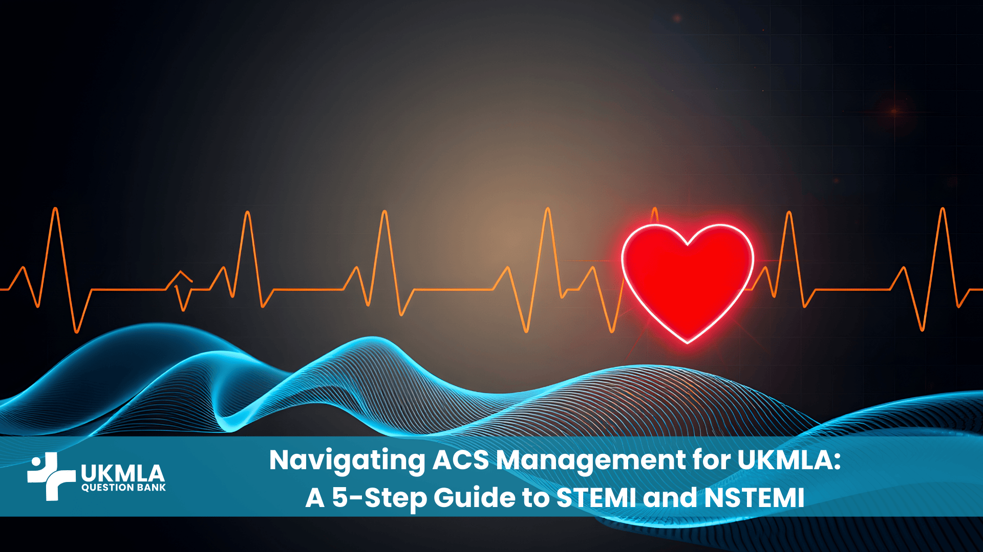
Mastering acs management ukmla protocols is arguably one of the most high-yield investments you can make for your exams. Whether you are facing a time-pressured Single Best Answer (SBA) question in the AKT or a frantic emergency scenario in the CPSA, chest pain is a universal presentation.
Acute Coronary Syndrome (ACS) covers a spectrum of conditions characterized by a sudden reduction in blood flow to the heart: ST-Elevation Myocardial Infarction (STEMI), Non-ST Elevation Myocardial Infarction (NSTEMI), and Unstable Angina. For the definitive UK standards, always refer to the NICE Guideline [NG185] on Acute Coronary Syndromes and the Resuscitation Council UK algorithms.
For a junior doctor or a medical student, the challenge isn’t just knowing the pathophysiology; it is about the speed of decision-making. You must rapidly differentiate between a patient who needs immediate catheterisation (PPCI) and one who requires medical stabilisation.
In this guide, we will break down the complex NICE guidelines and Resuscitation Council protocols into a logical, 5-step framework. We will cover everything from the initial “door-to-needle” decisions to the long-term pharmacology that frequently appears in exams.
Rapid Differentiation is Vital: Success depends on quickly distinguishing STEMI (ST-elevation) from NSTEMI/UA (ST-depression/T-wave inversion) via ECG.
Time is Muscle: For STEMI, the goal is reperfusion (PPCI) within 120 minutes of diagnosis.
Risk Stratification: NSTEMI management relies on the GRACE score to determine the timing of angiography.
The MONAC Protocol: Modern management involves Morphine, Oxygen (only if hypoxic), Nitrates, Aspirin (300mg), and a second antiplatelet (Clopidogrel/Ticagrelor).
Secondary Prevention: Remember the “6 As” for discharge planning—a favourite topic for AKT prescribing questions.
Step 1: Rapid Assessment and The Foundation
The initial assessment of a patient with chest pain is a race against time. In the UKMLA CPSA, you will likely be assessed on your ability to take a focused history while simultaneously initiating management. You cannot afford to be passive.
The SOCRATES Pain History
While you may know the mnemonic well, the application of SOCRATES in a cardiac context is specific. You are hunting for red flags that scream “Ischaemia”.
Character: Heaviness, crushing, tight band, “elephant on chest”.
Radiation: To the left arm, jaw, or neck (classic), or epigastrium (often confused with indigestion).
Associated Symptoms: Autonomic features are key predictors of infarction: sweating (diaphoresis), nausea, vomiting, and breathlessness.
Note: In diabetic patients and the elderly, “silent MIs” are common. They may present solely with shortness of breath, syncope, or confusion, without any chest pain.
Candidate: “Can you describe the pain for me? Does it feel like a weight or tightness?” Patient: “It feels heavy.” Candidate: “Does that pain move anywhere, like to your jaw or arm?” Patient: “My left arm hurts.” Candidate: “And have you felt sweaty or sick to your stomach since it started?”
Risk Factor Stratification
In the AKT, the clinical vignette will often list risk factors to nudge you toward a cardiac diagnosis. Group them mentally:
Table 1: ECG Lead Territories and Coronary Artery Correlation
Infarct Location
ECG Leads Affected
Coronary Artery Involved
Anterior
V1 – V4
Left Anterior Descending (LAD)
Lateral
I, aVL, V5, V6
Circumflex (LCx) or LAD
Inferior
II, III, aVF
Right Coronary Artery (RCA) – 80%
Posterior
Reciprocal depression V1-V3
RCA or Circumflex
Cardiac Biomarkers (Troponin)
High-sensitivity Troponin (hs-cTn) is the gold standard for detecting myocardial necrosis.
STEMI: Do not wait for Troponin results to treat. The ECG is diagnostic.
NSTEMI vs. Unstable Angina: This differentiation is purely biochemical. Both may have similar ECG changes.
NSTEMI: Elevated Troponin.
Unstable Angina: Normal Troponin (no cell death), but ischaemia is present.
Be aware of false positives. As discussed in Interpreting Clinical Data for UKMLA, Troponin can be raised in sepsis, pulmonary embolism (PE), and chronic renal failure. The key is the rise and fall pattern seen in ACS.
Other Bedside Investigations
Chest X-Ray (CXR): Crucial to rule out alternative diagnoses like pneumothorax or aortic dissection (widened mediastinum) before giving anticoagulants. Check our CXR Interpretation Guide for tips.
Blood Tests: FBC (anaemia aggravates ischaemia), U&Es (prior to contrast for angio), Glucose (hyperglycaemia is common), Lipid Profile.
Step 3: Initial Medical Management (The ‘Stabilisation’ Phase)
Once ACS is suspected, you initiate the “MONA” protocol—though modern guidelines have tweaked this significantly.
The ‘MONAC’ Protocol Revisited
In your exams, be precise with drug indications. Do not just throw oxygen at everyone.
M – Morphine: IV Morphine (e.g., 5-10mg) + an anti-emetic (Metoclopramide 10mg IV). Pain causes sympathetic surge, which increases heart rate and oxygen demand.
O – Oxygen:Only if saturation is <94%. There is strong evidence that hyperoxia can cause vasoconstriction and worsen infarct size.
N – Nitrates (GTN): Sublingual spray initially. If pain persists, start an IV GTN infusion.
Caution: Avoid in Inferior MI (RCA territory) if the patient is hypotensive, as they rely on preload.
Contraindication: Recent use of Sildenafil (Viagra).
A – Aspirin: 300mg stat (chewed or dispersible). This inhibits COX-1 and prevents further platelet aggregation.
C – Clopidogrel / Ticagrelor (The P2Y12 Inhibitor):
Most ACS patients now receive Ticagrelor (180mg loading) over Clopidogrel, unless they are at high bleeding risk or taking oral anticoagulants.
Prasugrel is often used in patients proceeding directly to PCI.
Anticoagulation
Anticoagulation prevents the thrombus from propagating.
NSTEMI:Fondaparinux (2.5mg SC daily) is the first line (unless angiography is immediate/urgent, then Unfractionated Heparin is used).
STEMI: Unfractionated Heparin is usually given in the cath lab during PCI.
Step 4: Definitive Management Pathways
This is where the road splits. Your management depends entirely on the ECG findings from Step 2.
STEMI Pathway: Time is Muscle
The coronary artery is completely occluded. It needs to be opened immediately.
Shutterstock
Primary PCI (PPCI): This is the gold standard. It involves angioplasty and stenting. It is indicated if it can be performed within 120 minutes of the time the diagnosis is made.
Thrombolysis (Fibrinolysis): Only used if PCI cannot be achieved within 120 minutes (e.g., rural setting). Tissue Plasminogen Activator (tPA) is used, followed by transfer to a PCI centre.
“What to Say” Script: Explaining PCI
Candidate: “We have identified a blockage in one of the arteries supplying your heart. The best treatment is a procedure called Primary PCI. We will take you to a specialist room, insert a small tube through your wrist or groin, and use a balloon and a metal mesh called a stent to open the artery and restore blood flow. This is the most effective way to limit damage to your heart.”
NSTEMI/UA Pathway: Risk Stratification
The artery is likely partially occluded. There is time to assess risk using the GRACE Score. This score calculates 6-month mortality risk based on age, HR, BP, Creatinine, and Troponin levels.
Aldosterone Antagonist: E.g., Eplerenone. Indicated if there is clinical heart failure or Left Ventricular Ejection Fraction (LVEF) <40%.
Lifestyle Modification
Driving: Patients cannot drive for 1 week (if successful angioplasty) or 4 weeks (if not successfully treated/conservative). They do not usually need to inform the DVLA for a standard Group 1 license unless they have disqualifying symptoms.
Smoking: Strict cessation advice.
Cardiac Rehab: All patients should be referred.
Clinical Scenarios: Putting it into Practice
Scenario 1: The ‘Indigestion’ that wasn’t
Patient: 55-year-old male, heavy smoker. Presentation: Burning epigastric pain for 2 hours, radiating to the jaw. He took Gaviscon with no relief. Vitals: HR 50, BP 90/60. ECG: ST elevation in II, III, and aVF.
Management: This is an Inferior STEMI (RCA territory). The bradycardia and hypotension confirm RCA involvement (supplying the SA/AV nodes).
Action: Activate PPCI lab immediately.
Caution: Avoid Nitrates and Morphine if BP is low; they reduce preload, which the right ventricle depends on. Give IV fluids if hypotensive. Give Aspirin 300mg and Ticagrelor 180mg.
Scenario 2: The Silent NSTEMI
Patient: 70-year-old female with Type 2 Diabetes. Presentation: “Just feeling weak and breathless” for 1 day. No chest pain. ECG: T-wave inversion in V1-V4. Troponin: Highly elevated (450 ng/L).
Management: This is an NSTEMI. The diabetes masked the pain.
Action: Admit to CCU. Calculate GRACE score. Start Fondaparinux 2.5mg SC, Aspirin 300mg, and Ticagrelor 180mg. Monitor for heart failure.
Practice Exercise: Test Your Knowledge
Question 1: A 62-year-old man is discharged 3 days after a STEMI managed with PPCI. He has no history of cough or renal failure. Which of the following medication combinations is most appropriate for discharge?
A) Aspirin, Clopidogrel, Simvastatin, Ramipril, Bisoprolol B) Aspirin, Ticagrelor, Atorvastatin, Ramipril, Bisoprolol C) Aspirin, Warfarin, Atorvastatin, Ramipril, Bisoprolol D) Clopidogrel only, Atorvastatin, Ramipril, Bisoprolol E) Aspirin, Ticagrelor, Atorvastatin, Furosemide
Correct Answer: B
Question 2: Which of the following patients requires urgent (<2 hours) angiography following an NSTEMI diagnosis?
A) A patient with a GRACE score of 2% B) A patient with ongoing chest pain despite maximal medical therapy C) A patient whose Troponin has peaked and is now falling D) A patient with diabetes mellitus E) A patient with T-wave inversion in lead III
Correct Answer: B
Frequently Asked Questions (FAQ) about ACS Management
No. NICE guidelines state oxygen should only be given if O2 sats are <94%. Hyperoxia can cause vasoconstriction, potentially worsening myocardial ischaemia. There is evidence suggesting routine oxygen in non-hypoxic patients may increase myocardial injury.
Usually, DAPT is continued for 12 months post-ACS. After 12 months, the patient typically steps down to Aspirin monotherapy lifelong. In patients with very high bleeding risk, this duration might be shortened to 3-6 months under specialist guidance.
Both can present with similar symptoms and ECG changes (ST depression/T-inversion). The difference is Troponin. NSTEMI has elevated Troponin (indicating cell death); Unstable Angina has normal Troponin. Essentially, Unstable Angina is ischaemia without infarction.
Thrombolysis is only used for STEMI if PPCI cannot be performed within 120 minutes of the diagnosis (e.g., remote locations). It involves giving a “clot-buster” drug IV. If thrombolysis is successful, the patient is still usually transferred for angiography, but not as an immediate “primary” procedure.
Inferior MIs often involve the Right Coronary Artery, which supplies the Right Ventricle. The RV is preload-dependent. Nitrates dilate veins and reduce preload, which can cause severe hypotension in these patients. This can lead to haemodynamic collapse.
New (or presumed new) Left Bundle Branch Block (LBBB) in the context of chest pain should be treated as a STEMI equivalent. It masks ST changes, so you cannot rule out a STEMI. Guidelines recommend initiating the PPCI pathway immediately.
They must stop driving for at least 1 week (if successful angioplasty) or 4 weeks (if not). They do not need to inform the DVLA for a standard car license unless symptoms persist. Bus/Lorry drivers have stricter rules (6 weeks off + testing).
For secondary prevention of cardiovascular disease (ACS), the dose is 80mg ON (high intensity), regardless of the cholesterol level. This high dose has pleiotropic effects, stabilising plaques beyond just lowering lipids.
This is a delayed pericarditis that occurs 2-6 weeks after an MI. It presents with pleuritic chest pain, fever, and a raised ESR. It is an autoimmune reaction to heart muscle neo-antigens and is treated with NSAIDs or Colchicine.
Beta-blockers (like Bisoprolol) reduce heart rate and contractility, thereby reducing myocardial oxygen demand and preventing arrhythmias. They have been proven to reduce long-term mortality and prevent remodelling of the heart muscle.
Conclusion
Managing acs management ukmla topics effectively requires a blend of sharp diagnostic skills and pharmacological precision. Remember, the examiner wants to see that you are safe. This means you recognise the STEMI immediately, you do not delay the call to the cath lab, and you do not discharge a patient without the life-prolonging “6 As”.
Whether it is distinguishing a subtle NSTEMI on an ECG or explaining the importance of DAPT to a patient in the CPSA, following this 5-step structure will ensure you cover all bases.
Your Next Steps
Review ECG Territories: Go back to the table in Step 2 and memorise which leads correspond to which artery.
Practice Prescribing: Write out a mock drug chart for a post-MI patient, ensuring you get the doses of Atorvastatin (80mg) and Aspirin (75mg) correct.
Roleplay: Find a study partner and practice the “Explanation of PCI” script until it flows naturally.