Introduction
Deep Vein Thrombosis (DVT) is one of the most common acute medical presentations you will encounter as a junior doctor. The stakes are high: miss a DVT, and the patient could suffer a fatal Pulmonary Embolism (PE). Conversely, treat a patient incorrectly, and you risk a major bleed. For a general overview of the condition and patient information, the NHS Deep Vein Thrombosis Guide is a useful reference.
However, for the UKMLA, the diagnostic pathway is rigid. You cannot simply “eyeball” a swollen leg. You must apply the specific dvt diagnosis uk algorithm mandated by NICE Guideline [NG158]. Whether it is sequencing tests in the AKT or explaining the need for a scan to a patient in the CPSA, precision is key.
In this guide, we will break down the NICE pathway into 5 logical steps, covering the Wells Score calculation, the critical “4-hour rule,” and the modern use of Direct Oral Anticoagulants (DOACs).
Table of Contents
ToggleKey Takeaways
The Two-Level Wells Score: Distinguishing between “DVT Likely” (2 points or more) and “DVT Unlikely” (1 point or less) is the first critical step.
The “4-Hour Rule”: If a scan cannot be obtained within 4 hours for a “Likely” patient, you must start interim anticoagulation.
D-Dimer Utility: Only useful in the “Unlikely” pathway to rule out DVT. It has no diagnostic value in the “Likely” pathway.
First-Line Treatment: DOACs (Apixaban or Rivaroxaban) are now standard first-line therapy, replacing LMWH/Warfarin for most patients.
Investigation Sequence: Leg Ultrasound (US) is the gold standard, but knowing what to do when it’s negative (repeat scan?) is a key exam discriminator.
Step 1: Clinical Assessment and The Wells Score
The classic presentation is a unilateral swollen, tender calf. However, clinical judgment alone is unreliable. You must use the Two-Level DVT Wells Score to risk-stratify the patient.
The Two-Level DVT Wells Score
You calculate the score based on the following criteria.
| Clinical Feature | Points |
|---|---|
| Active cancer (treatment ongoing, within 6 months, or palliative) | +1 |
| Paralysis, paresis, or recent plaster immobilisation of lower extremities | +1 |
| Recently bedridden >3 days or major surgery within 12 weeks | +1 |
| Localised tenderness along the distribution of the deep venous system | +1 |
| Entire leg swollen | +1 |
| Calf swelling >3 cm larger than asymptomatic side (measured 10 cm below tibial tuberosity) | +1 |
| Pitting oedema confined to the symptomatic leg | +1 |
| Collateral superficial veins (non-varicose) | +1 |
| Previously documented DVT | +1 |
| Alternative diagnosis at least as likely as DVT | -2 |
Score ≥ 2: DVT Likely
Score ≤ 1: DVT Unlikely
Step 2: The ‘DVT Likely’ Pathway (Score ≥2)
If the patient scores 2 or more, the probability of DVT is high. Do not waste time with a D-dimer yet; go straight for imaging.
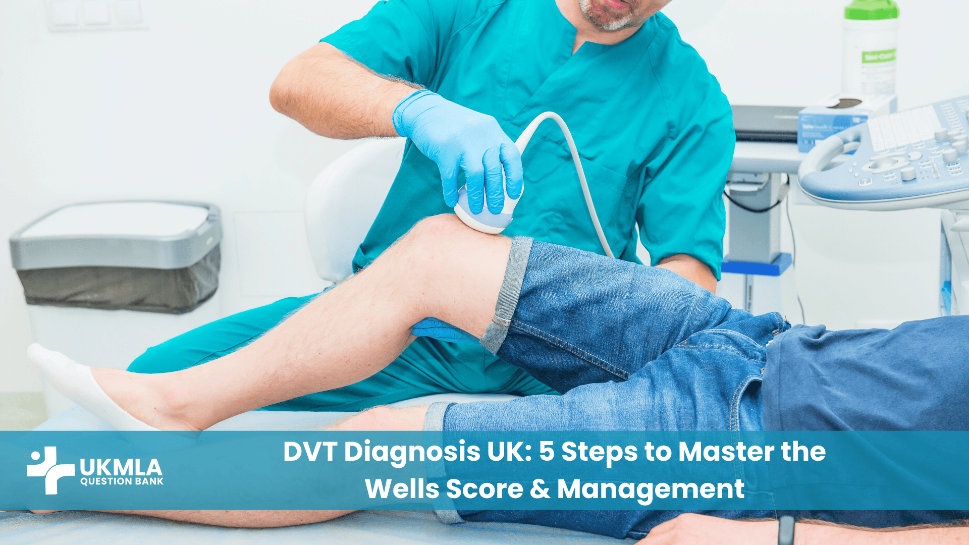
Immediate Action: Proximal Leg Vein Ultrasound
Ideally, you should obtain a proximal leg vein ultrasound scan within 4 hours of the request.
The ‘Interim’ Rule
This is a favourite AKT question trap.
Scenario: It is 8:00 PM on a Friday. The ultrasound department is closed until morning.
Action: You must start Interim Therapeutic Anticoagulation immediately. Do not leave the patient untreated overnight.
Drug of Choice: Usually Apixaban or Rivaroxaban (unless contraindicated, e.g., in pregnancy or renal failure, where LMWH is used).
Review: Perform the scan within 24 hours.
Negative Scan?
If the scan is negative but the D-dimer is raised, there is still a risk of a distal DVT extending.
Action: Stop anticoagulation but arrange a repeat scan in 6–8 days.
Step 3: The ‘DVT Unlikely’ Pathway (Score ≤1)
If the score is 0 or 1, the probability is low. You need a rule-out test.
The Gatekeeper: D-dimer Test
Perform a quantitative D-dimer blood test. For a refresher on interpreting this, see our guide on Master Interpreting Clinical Data.
Negative D-dimer: DVT is excluded. You do not need an ultrasound. Consider alternative diagnoses like Baker’s cyst, cellulitis, or muscle injury.
Positive D-dimer: DVT is possible. Proceed to Ultrasound.
Apply the same “4-hour rule” as above. If the scan is delayed >4 hours, start interim anticoagulation.
Step 4: Definitive Management (Anticoagulation)
Once DVT is confirmed, long-term anticoagulation is required. Before starting, check baseline bloods: FBC, U&Es, LFTs, and a Coagulation Screen.
First Line: DOACs
Apixaban or Rivaroxaban are the first-line treatments for most patients.
Advantage: No monitoring (INR) required, oral administration.
Apixaban Dosing: 10mg BD for 7 days, then 5mg BD.
Rivaroxaban Dosing: 15mg BD for 21 days, then 20mg OD.
Second Line: LMWH followed by DOAC
If neither Apixaban nor Rivaroxaban is suitable, or if lead-in is required for Edoxaban/Dabigatran:
Start LMWH (e.g., Enoxaparin) for at least 5 days.
Switch to Edoxaban or Dabigatran thereafter.
Renal Impairment
If the patient has severe renal impairment (Creatinine Clearance < 15 ml/min), DOACs are generally contraindicated.
Action: Use LMWH (dose adjusted) or Unfractionated Heparin. Refer to our Ultimate Guide to UK Blood Tests for monitoring renal function.
Step 5: Duration and Follow-up
How long do you treat for? This depends on the cause.
Provoked DVT: (e.g., surgery, trauma, long-haul flight). The risk factor is transient.
Duration: 3 months is usually sufficient.
Unprovoked DVT: No obvious cause found. This suggests a pro-thrombotic tendency or hidden malignancy.
Duration: 3 months initially, then review. If bleeding risk is low, consider continuing long-term (lifelong).
Cancer-Associated Thrombosis:
Duration: 6 months to lifelong (while cancer is active).
Drug: DOACs (Edoxaban) are now often used, except in gastrointestinal cancers where LMWH is preferred due to bleeding risk.
Clinical Scenarios: Putting it into Practice
Scenario 1: The Late Friday Admission
Patient: 45-year-old male, calf pain after returning from Australia. Wells Score = 4 (Likely).
Situation: It is 10 PM. Ultrasound is unavailable until 9 AM.
Management: Do not wait. Prescribe Apixaban 10mg PO stat. Book the scan for tomorrow morning. This is the “interim anticoagulation” rule in action.
Scenario 2: The Confusing Negative
Patient: 60-year-old female. Wells Score = 2 (Likely).
Investigation: Ultrasound shows no DVT. However, her D-dimer is significantly raised.
Management: You cannot rule out DVT yet. A distal clot might be too small to see or might extend later. Stop the Apixaban for now, but bring her back for a repeat ultrasound in 6-8 days.
Practice Exercise: Test Your Knowledge
Question 1: A 32-year-old woman (12 weeks pregnant) presents with a swollen left leg. A DVT is suspected. What is the most appropriate initial investigation?
A) D-dimer blood test
B) CT Pulmonary Angiogram (CTPA)
C) Compression Duplex Ultrasound of the leg
D) Venogram
E) Wells Score calculation
Correct Answer: C
Question 2: A 65-year-old man is diagnosed with a proximal DVT. He has a history of Stage 4 Chronic Kidney Disease with an eGFR of 12 ml/min. Which of the following is the most appropriate initial anticoagulation?
A) Apixaban
B) Rivaroxaban
C) Warfarin
D) Unfractionated Heparin (or adjusted dose LMWH)
E) Dabigatran
Correct Answer: D
Frequently Asked Questions (FAQ) about DVT Diagnosis
No. The standard Wells Score is not validated for use in pregnancy. Pregnant patients with suspected DVT should generally proceed directly to compression ultrasound.
This provides a standardised landmark to ensure reproducibility. Measuring at random points can lead to false positives due to muscle bulk differences.
Proximal DVT involves the popliteal vein or above (femoral, iliac). These carry a high risk of PE. Distal DVT is below the knee (calf veins). These have a lower risk of PE but can extend proximally.
In any “Unprovoked” DVT (no surgery, travel, or trauma) in a patient >40 years old. NICE recommends offering abdominal/pelvic CT and mammogram/chest X-ray to screen for occult malignancy.
No. Antiplatelets like Aspirin are ineffective for treating venous thrombosis. They work on platelets (arterial clots), whereas DVT requires anticoagulants (fibrin pathway).
The treatment is essentially the same (anticoagulation), but the urgency and monitoring (oxygen, haemodynamics) will be dictated by the PE severity.
Generally no. Cancer often elevates D-dimer regardless of thrombosis, leading to false positives. Most guidelines suggest going straight to imaging for cancer patients.
A rare, severe form of DVT where massive obstruction causes ischaemia (blue leg) and can lead to gangrene. This is a surgical emergency requiring thrombolysis or thrombectomy.
Yes. Most stable patients with DVT are treated as outpatients. Admission is only needed if there is severe pain, comorbidity, high bleeding risk, or suspicion of PE.
It acts as a filter. We only use D-dimer if the Wells Score is “Unlikely” (≤1). If the score is “Likely” (≥2), the pre-test probability is too high, and a negative D-dimer might be a false negative, so we skip it.
Conclusion
Mastering dvt diagnosis uk protocols is about discipline. It is tempting to jump straight to a scan or a blood test, but the NICE Guideline [NG158] demands a structured approach starting with the Wells Score.
Remember the golden rules for your exam:
Calculate: Wells Score first.
Stratify: Likely (Scan) vs. Unlikely (D-dimer).
Act: If the scan is delayed >4 hours in a likely patient, treat immediately.
Your Next Steps
Memorise the Score: You don’t need to know every point value perfectly, but know the big ones: Active Cancer, Swelling >3cm, and Bedridden/Surgery.
Check the Drug Chart: In your next clinical placement, look at the drug chart of a DVT patient. Note the dosing of Apixaban (Loading dose vs Maintenance).
Practice Explaining: Try explaining to a friend why they need a scan even though their D-dimer was normal (or vice versa) to prepare for the CPSA.