Introduction
Neurology is a cornerstone of medical practice, challenging yet immensely rewarding. For medical students and international medical graduates preparing for the UK Medical Licensing Assessment (UKMLA), particularly the Applied Knowledge Test (AKT), a robust understanding of neurological conditions and diagnostic pathways is paramount. This comprehensive guide provides UKMLA Neurology Essentials, delving into 7 high-yield syndromes frequently encountered in clinical practice and heavily tested in the AKT, alongside effective diagnostic approaches to ensure your success.
Table of Contents
Toggle
Why Neurology is Crucial for Your UKMLA Success
Neurology constitutes a significant domain within the UKMLA, reflecting its prevalence and critical importance in patient care. The AKT assesses not just your factual knowledge but your capacity for clinical reasoning and your ability to manage patients safely and effectively. This makes a deep dive into UKMLA Neurology Essentials indispensable.
The UKMLA AKT’s Emphasis on Clinical Reasoning
The AKT is designed to evaluate your application of medical knowledge to real-world scenarios. In neurology, this means interpreting complex patient vignettes, localizing lesions, selecting appropriate investigations, and formulating initial management plans. Questions often present subtle clues, requiring a systematic approach to diagnosis that mirrors actual clinical practice. Developing this skill is a central part of mastering your UKMLA Neurology Essentials.
The Importance of a Structured Diagnostic Approach
Given the intricate nature of the nervous system and the overlap in symptoms, a structured approach is fundamental. Without a methodical framework, crucial diagnostic information can be overlooked, potentially leading to misdiagnosis or delayed treatment. A systematic process, moving from history to examination and then to investigations, ensures thoroughness and accuracy, which are highly valued in both the UKMLA and your future medical career.
To gain a broader understanding of how neurology fits into the overall UKMLA framework, it’s beneficial to consult official resources. You can explore The GMC UKMLA Content Map: Your Blueprint for Success to see how various topics are weighted and connected.
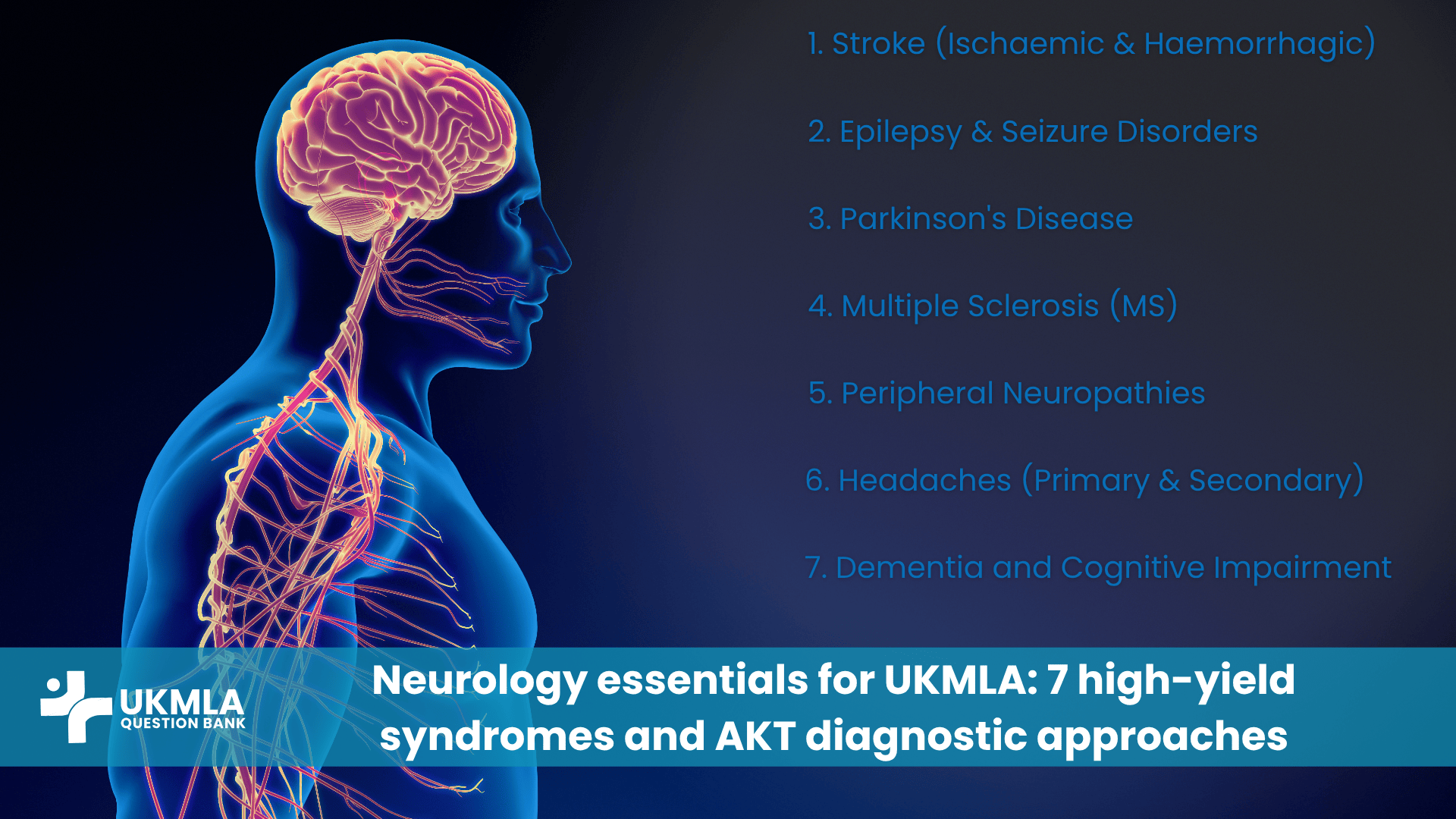
UKMLA Neurology Essentials: The 7 High-Yield Syndromes
This section outlines seven neurological conditions that consistently appear in the UKMLA, providing you with the UKMLA Neurology Essentials for each.
1. Stroke (Ischaemic & Haemorrhagic)
Stroke is a medical emergency demanding rapid recognition and intervention. The UKMLA frequently tests its acute presentation and management.
- Key Diagnostic Features: Sudden onset of focal neurological deficit. Ischaemic stroke (85%) results from vessel occlusion, presenting with symptoms based on the affected artery (e.g., Middle Cerebral Artery: contralateral weakness/aphasia; Posterior Cerebral Artery: visual field defects). Haemorrhagic stroke (15%) often presents with severe headache, vomiting, and altered consciousness due to bleeding, often linked to uncontrolled hypertension or aneurysm rupture.
- Initial Assessment: The FAST (Face, Arm, Speech, Time) test is crucial for rapid identification. Urgent non-contrast CT head is mandatory to differentiate ischaemic from haemorrhagic, as this distinction dictates immediate treatment.
- Differential Diagnoses: Seizures (especially Todd’s paralysis, a post-ictal focal weakness), migraine with aura, hypoglycaemia, Bell’s palsy (for isolated facial droop).
- Brief Management Principles: For eligible ischaemic strokes, thrombolysis (alteplase) within 4.5 hours and/or mechanical thrombectomy (within 6-24 hours) may be indicated. For haemorrhagic stroke, strict blood pressure control and reversal of anticoagulation are vital, with neurosurgical consultation for appropriate cases. General supportive care (ABC, glucose, temperature) applies to all stroke patients. Please follow the official guidelines on Stroke and transient Ischaemic attache here.
2. Epilepsy & Seizure Disorders
Epilepsy is defined by recurrent, unprovoked seizures. The AKT assesses classification, diagnostic workup, and acute/long-term management.
- Key Diagnostic Features & Classification: A seizure is a transient occurrence of signs and/or symptoms due to abnormal excessive or synchronous neuronal activity in the brain. Epilepsy is diagnosed after two or more unprovoked seizures (>24 hours apart), or one unprovoked seizure with a high risk of recurrence. Seizures are broadly classified by onset: Focal onset (symptoms correlate with a specific brain region, can be aware or impaired awareness) and Generalized onset (affecting both hemispheres simultaneously, e.g., tonic-clonic, absence, myoclonic, atonic).
- Diagnostic Workup: A detailed history is paramount, especially from a witness, to differentiate epileptic seizures from non-epileptic events (e.g., syncope, psychogenic non-epileptic seizures). An EEG (Electroencephalogram) helps classify seizure type and localize seizure onset, though a normal EEG doesn’t rule out epilepsy. MRI brain with an epilepsy protocol is essential to identify structural causes (e.g., tumours, malformations, hippocampal sclerosis). Blood tests rule out metabolic causes (e.g., hypoglycaemia, electrolyte imbalances).
- Differential Diagnoses: Syncope (especially vasovagal), psychogenic non-epileptic seizures (PNES), transient ischaemic attacks (TIAs), cardiac arrhythmias.
- Brief Acute & Long-Term Management: For acute seizures, ensure patient safety and ABCs; if prolonged (>5 minutes or recurrent), administer benzodiazepines (e.g., IV lorazepam or buccal midazolam). Long-term management involves Antiepileptic Drugs (AEDs) chosen based on seizure type, comorbidities, and side effect profiles (e.g., sodium valproate, lamotrigine, levetiracetam). Patient education on driving restrictions and avoiding triggers is also important.
3. Parkinson’s Disease
A progressive neurodegenerative disorder affecting dopamine neurons in the substantia nigra, often tested for its classic motor and non-motor symptoms.
- Cardinal Symptoms (TRAP): These are the core motor features: Tremor (resting, typically “pill-rolling,” often asymmetrical), Rigidity (increased resistance to passive movement, often “cogwheel” rigidity), Akinesia/Bradykinesia (slowness and reduction in amplitude of movement, masked facies, reduced arm swing), and Postural Instability (impaired balance, leading to falls, usually a later feature). Other non-motor symptoms are very common and can precede motor symptoms: anosmia (loss of smell), constipation, REM sleep behaviour disorder, depression, anxiety, and cognitive impairment.
- Diagnostic Criteria: Diagnosis is primarily clinical, based on the presence of bradykinesia plus at least one of resting tremor or rigidity. Asymmetry of motor symptoms is a common early sign. DaTscan (Dopamine Transporter Scan) can aid differentiation from mimics in ambiguous cases (showing reduced striatal dopamine transporter uptake in PD).
- Differential Diagnoses: Essential tremor (action tremor), drug-induced parkinsonism (e.g., from antipsychotics), atypical parkinsonian syndromes (e.g., Multiple System Atrophy, Progressive Supranuclear Palsy).
- Brief Pharmacological Management Principles: Levodopa (often combined with carbidopa or benserazide) is the most effective symptomatic treatment. Dopamine agonists (e.g., ropinirole, pramipexole) may be used as initial therapy, especially in younger patients, to delay levodopa-related motor complications. MAO-B inhibitors (e.g., selegiline, rasagiline) and COMT inhibitors (e.g., entacapone) are also used. Non-pharmacological therapies like physiotherapy, occupational therapy, and speech therapy are crucial for maintaining function.
4. Multiple Sclerosis (MS)
A chronic inflammatory demyelinating disease of the central nervous system (CNS) with highly variable presentations, reflecting lesions in different areas of the brain and spinal cord.
- Common Presentations: MS often presents with discrete relapses (new or worsening neurological symptoms lasting >24 hours, in the absence of fever/infection) followed by remission. Common symptoms include optic neuritis (painful monocular vision loss, often with colour desaturation), sensory symptoms (numbness, tingling, Lhermitte’s sign – an electric shock sensation down the spine with neck flexion), motor weakness, spasticity, ataxia, diplopia (double vision due to ocular motor nerve involvement), and debilitating fatigue.
- Diagnostic Criteria: Diagnosis is based on the McDonald Criteria, requiring evidence of dissemination in space (lesions in at least two different CNS areas) and dissemination in time (evidence of lesions occurring at different times, or a clinical relapse plus a new MRI lesion). MRI brain and spinal cord is the primary diagnostic tool, showing characteristic white matter lesions. Lumbar Puncture (LP) for analysis of cerebrospinal fluid (CSF) can show oligoclonal bands (OCBs), which are highly suggestive of MS, but not always mandatory for diagnosis if MRI criteria are met.
- Differential Diagnoses: Other inflammatory/demyelinating conditions (e.g., neuromyelitis optica spectrum disorder), B12 deficiency, vasculitis, sarcoidosis, stroke mimics.
- Brief Management (Acute Relapse & DMTs): Acute relapses are typically managed with a short course of high-dose corticosteroids (e.g., methylprednisolone) to shorten relapse duration and severity. Disease-Modifying Therapies (DMTs) are used long-term to reduce relapse frequency, severity, and slow disease progression (e.g., interferon beta, glatiramer acetate, natalizumab, ocrelizumab). Symptomatic management for fatigue, spasticity, bladder dysfunction, and pain is also vital.
5. Peripheral Neuropathies (e.g., Diabetic Neuropathy, GBS)
Peripheral neuropathies are a group of disorders affecting nerves outside the brain and spinal cord, causing sensory, motor, or autonomic symptoms depending on the nerve fibres involved.
- Common Causes: Diabetic Neuropathy is the most prevalent cause, typically presenting with a “glove and stocking” distribution of sensory loss, often with pain, numbness, or tingling. Guillain-Barré Syndrome (GBS) is an acute, rapidly progressive, ascending flaccid paralysis often triggered by an infection (e.g., Campylobacter jejuni) and is a medical emergency. Other causes include alcohol abuse, vitamin deficiencies (B1, B12), uraemia (kidney failure), hypothyroidism, certain drugs (e.g., chemotherapy agents), autoimmune diseases, and entrapment neuropathies (e.g., carpal tunnel syndrome).
- Patterns of Presentation: Neuropathies can present in various patterns: Mononeuropathy (affecting a single nerve, e.g., wrist drop in radial neuropathy), Mononeuropathy multiplex (affecting multiple isolated nerves), Polyneuropathy (symmetrical, widespread, often length-dependent, starting distally and progressing proximally), and Radiculopathy (nerve root involvement, e.g., dermatomal sensory loss, myotomal weakness).
- Key Diagnostic Tests: Nerve Conduction Studies (NCS) and Electromyography (EMG) are the gold standard for confirming neuropathy, classifying it (axonal vs. demyelinating), and assessing severity and distribution. Blood tests are crucial to identify underlying systemic causes (e.g., HbA1c for diabetes, B12 levels, thyroid function tests, autoimmune antibodies). Lumbar Puncture (LP) for CSF analysis is characteristic in GBS (elevated protein with normal cell count – albuminocytological dissociation).
- Brief Management Strategies: Treatment focuses on addressing the underlying cause (e.g., strict glycemic control in diabetes). Symptomatic treatment for neuropathic pain often involves medications like gabapentin or pregabalin. Physiotherapy and occupational therapy are vital for managing motor weakness. Specific treatment for GBS involves IV immunoglobulin (IVIg) or plasma exchange.
6. Headaches (Primary & Secondary)
Headaches are among the most common neurological complaints, and the UKMLA often assesses your ability to differentiate benign primary headaches from dangerous secondary causes that require urgent investigation.
- Key Diagnostic Features:
- Primary Headaches:
- Migraine: Typically unilateral, throbbing, moderate-to-severe, often associated with nausea, photophobia, and phonophobia. May have an aura (visual, sensory, or motor disturbances preceding the headache).
- Tension-Type Headache: Bilateral, pressing/tightening (non-pulsating), mild-to-moderate intensity, not aggravated by routine physical activity. No nausea/vomiting, and either photophobia or phonophobia (but not both).
- Cluster Headache: Severe, strictly unilateral pain, usually orbital, supraorbital, or temporal, lasting 15-180 minutes, occurring in “clusters.” Associated with ipsilateral autonomic symptoms (e.g., lacrimation, conjunctival injection, nasal congestion, ptosis, miosis, facial sweating).
- Secondary Headaches (Red Flags indicating serious underlying pathology):
- Sudden onset “thunderclap” headache (reaching maximum intensity within one minute) – highly suggestive of Subarachnoid Haemorrhage (SAH).
- Worst headache of life.
- Fever, neck stiffness, photophobia (suggesting Meningitis).
- Focal neurological deficits (e.g., weakness, numbness, visual changes).
- Papilledema (swelling of the optic disc).
- Headache awakened from sleep.
- New headache onset in patients >50 years old (consider Giant Cell Arteritis).
- History of head trauma, cancer, or immunosuppression.
- Primary Headaches:
- Diagnostic Workup: Primarily relies on a detailed history. If red flags are present, urgent investigations are required: CT head (non-contrast) to rule out acute haemorrhage (e.g., SAH) or mass lesions. If SAH is suspected but CT is negative, a Lumbar Puncture (LP) for xanthochromia (yellow discoloration of CSF due to breakdown products of blood) is needed. Further imaging (e.g., MRI, MR venography) may be indicated based on suspected cause.
- Brief Management Principles: For primary headaches, management involves abortive treatments (e.g., triptans, NSAIDs for migraine; simple analgesia for tension) and prophylactic treatments (e.g., beta-blockers for migraine; verapamil for cluster). For secondary headaches, immediate investigation and targeted treatment of the underlying cause are paramount.
7. Dementia and Cognitive Impairment
Dementia refers to a progressive decline in cognitive function (e.g., memory, language, executive function, visuospatial skills) severe enough to interfere with daily activities. The UKMLA assesses its common causes and diagnostic pathways.
- Key Diagnostic Features: Progressive decline in multiple cognitive domains.
- Alzheimer’s Disease: Most common cause. Characterized by insidious onset and gradual progression, with early and prominent memory impairment, followed by difficulties in language, visuospatial skills, and executive function.
- Vascular Dementia: Often presents with a more step-wise cognitive decline, commonly associated with a history of stroke or cardiovascular risk factors, and may have focal neurological signs.
- Lewy Body Dementia (LBD): Characterized by fluctuating cognition (variations in attention and alertness), recurrent well-formed visual hallucinations, and spontaneous parkinsonism (often occurring at or before the onset of cognitive decline).
- Frontotemporal Dementia (FTD): Typically presents with earlier and more prominent changes in personality, behaviour (e.g., disinhibition, apathy), or language (e.g., progressive non-fluent aphasia, semantic dementia) compared to memory impairment.
- Diagnostic Workup: A thorough history (from both the patient and a reliable collateral source like a family member) is crucial to establish the pattern and impact of cognitive decline. Standardized cognitive assessments (e.g., MMSE, MoCA) provide objective measures. Blood tests are essential to rule out reversible causes of cognitive impairment (e.g., B12 deficiency, hypothyroidism, syphilis, electrolyte imbalances, renal/hepatic dysfunction). Structural neuroimaging (MRI brain) is preferred to assess for patterns of atrophy, vascular changes, and to rule out other treatable causes like brain tumours or normal pressure hydrocephalus.
- Differential Diagnoses: Delirium (acute onset, fluctuating, usually underlying medical cause), depression (can mimic dementia – “pseudodementia”), normal pressure hydrocephalus (triad of gait disturbance, urinary incontinence, and dementia), B12 deficiency, hypothyroidism, medication side effects.
- Brief Management Principles: Currently, there’s no cure for most forms of dementia. Management focuses on symptomatic treatment (e.g., cholinesterase inhibitors like donepezil for Alzheimer’s, LBD), managing behavioural and psychological symptoms (agitation, depression), providing support for carers, and addressing any reversible causes identified. A multidisciplinary team approach (including occupational therapy, speech therapy, social workers) is crucial.
Key Diagnostic Approaches for UKMLA Neurology
Beyond specific conditions, mastering systematic diagnostic principles is key to success in UKMLA Neurology Essentials.
Systematic History Taking in Neurology
A thorough history is often the single most powerful diagnostic tool in neurology, providing crucial clues to localize the problem and narrow the differential diagnosis.
- Onset and Progression: Was the onset sudden (e.g., stroke), acute (e.g., Guillain-Barré Syndrome, encephalitis), or insidious (e.g., neurodegenerative disease)? Is the course progressive, relapsing-remitting (e.g., Multiple Sclerosis), or static?
- Symptom Characterization: Obtain a precise and detailed description of the patient’s symptoms. Is it weakness (proximal/distal, focal/generalized), numbness (tingling, burning, pins and needles), dizziness (true vertigo vs. lightheadedness/presyncope), visual changes (vision loss, double vision), speech difficulties (dysarthria, aphasia), or balance issues?
- Associated Symptoms: Always ask about accompanying symptoms such as headache, fever, altered consciousness, bladder/bowel dysfunction, cognitive changes, and any systemic symptoms (e.g., rashes, joint pain) that might suggest a systemic cause.
- Red Flags: Actively probe for “red flags” that indicate serious or urgent pathology. These include thunderclap headache, rapid progression of symptoms, new-onset seizures in an adult without prior history, neck stiffness, photophobia, papilledema, or symptoms in the context of cancer or immunosuppression.
- Past Medical History, Medications, and Family History: These provide vital context. Ask about conditions like diabetes, hypertension, autoimmune diseases, recent infections, and any neurotoxic medications. A family history of neurological disorders can be significant.
Essential Neurological Examination Findings & Interpretation
The neurological examination systematically assesses the central and peripheral nervous systems, helping to confirm historical findings and precisely localize the lesion.
- Mental State: Assess the patient’s level of consciousness (using the Glasgow Coma Scale – GCS), orientation (time, place, person), and basic cognitive functions (e.g., attention, language, memory).
- Cranial Nerves: Systematically test all 12 cranial nerves (e.g., visual acuity/fields, pupillary reflexes, eye movements, facial sensation/movement, hearing, swallowing, speech, tongue movement). Deficits can localize lesions to the brainstem, specific nerves, or even higher cortical centres.
- Motor System: Begin with inspection (look for muscle wasting, fasciculations). Then assess muscle tone (spasticity, rigidity, flaccidity). Test muscle power in key movements using the Medical Research Council (MRC) scale (0-5). Elicit deep tendon reflexes (biceps, triceps, knee, ankle) and plantar reflexes. Distinguish between Upper Motor Neuron (UMN) signs (spasticity, hyperreflexia, Babinski sign) and Lower Motor Neuron (LMN) signs (flaccidity, hyporeflexia, muscle wasting, fasciculations).
- Sensory System: Test light touch, pain (pinprick), temperature, vibration (tuning fork), and proprioception (joint position sense). Note the pattern of sensory loss (e.g., dermatomal for radiculopathy, glove-and-stocking for polyneuropathy, sensory level for spinal cord lesions).
- Coordination & Gait: Assess cerebellar function with tests like finger-nose test, heel-to-shin test, and rapid alternating movements. Observe gait for specific patterns indicative of neurological pathology (e.g., ataxic, spastic, parkinsonian, foot drop). Perform Romberg’s test for proprioceptive loss.
- Interpretation: Synthesize all examination findings with the history to localize the lesion to the brain (cerebrum, cerebellum, brainstem), spinal cord, nerve root, plexus, peripheral nerve, neuromuscular junction, or muscle.
Interpreting Neuroimaging (CT/MRI Head)
Understanding the indications for and basic interpretation of neuroimaging is a key skill tested in the AKT.
- CT Head (Computed Tomography):
- Indications: Primarily used in acute settings: acute stroke (to rapidly rule out haemorrhage), head trauma, acute severe headache with red flags (e.g., suspected SAH), or suspected hydrocephalus.
- Key Findings: Acute haemorrhage appears as bright (hyperdense) areas. Early ischaemic changes can be subtle (sulcal effacement, loss of grey-white differentiation) but later appear as hypodensities. Can identify large mass lesions (tumours, abscesses) and fractures.
- MRI Head (Magnetic Resonance Imaging):
- Indications: Offers superior soft tissue contrast for more detailed imaging in non-acute settings or after negative CT: suspected Multiple Sclerosis (shows characteristic white matter plaques), Epilepsy (to identify subtle structural causes like hippocampal sclerosis), brain tumours, inflammatory conditions, subtle or chronic stroke, and detailed assessment of the posterior fossa, brainstem, and spinal cord.
- Key Findings: Excellent for white matter lesions, better for differentiating acute vs. chronic ischaemia, and identifying subtle or small tumours.
Table 1: Common Neurological Symptoms & Their High-Yield Causes for UKMLA
| Symptom | Key Differential Diagnoses (High-Yield for UKMLA) | Key Diagnostic Clues (History/Exam) |
|---|---|---|
| Headache | Migraine, Tension headache, Cluster headache, Subarachnoid haemorrhage, Meningitis, Brain tumour, Temporal arteritis | Onset, character, severity, associated symptoms (photophobia, fever, focal neurology), red flags (thunderclap, worst ever). |
| Weakness | Stroke, Spinal cord lesion, Peripheral neuropathy, Myasthenia gravis, GBS, MS, Motor neuron disease | Onset (acute/gradual), pattern (focal, hemi-, para-, tetra-, proximal/distal), associated sensory changes, bulbar symptoms. |
| Dizziness/Vertigo | Benign Paroxysmal Positional Vertigo (BPPV), Meniere’s disease, Vestibular neuritis, Stroke (posterior circulation) | True spinning sensation (vertigo), triggers (head position), associated hearing loss/tinnitus, focal neurology (central cause). |
| Vision Loss | Optic neuritis (MS), Stroke (posterior circulation), Retinal detachment, Glaucoma | Onset (sudden/gradual), painful/painless, monocular/binocular, field defect pattern, associated neurological symptoms. |
| Numbness/Tingling | Peripheral neuropathy (e.g., diabetic), Stroke, Spinal cord compression, MS, Radiculopathy | Pattern of distribution (dermatomal, glove-and-stocking), onset, progression, associated weakness/pain. |
Mastering AKT Questions: Applying Your Diagnostic Knowledge
Knowing the UKMLA Neurology Essentials is foundational, but applying this knowledge effectively under exam conditions is where you’ll shine.
Strategy for Single Best Answer (SBA) Questions
The majority of AKT questions are SBAs, requiring you to select the single best answer from five options.
- Read the Stem Carefully: Don’t skim. Identify the patient’s age, gender, presenting complaint, duration, key positive findings, and any relevant negatives. Pay close attention to “red flags” – these are often the quickest way to narrow down differentials.
- Identify the Core Question: Be clear about what is being asked. Is it a diagnosis? The next best investigation? Immediate management? A prognostic statement?
- Generate Differentials: Based on the information in the stem, quickly consider the most likely diagnoses. Think broadly first, then narrow down using specifics.
- Evaluate Options Systematically: Rule out options that are clearly incorrect or inconsistent with the stem. For the remaining options, compare them rigorously against all details provided, selecting the best answer, even if multiple options seem partially correct.
- Look for Distractors: Be aware of options that are plausible but don’t fit all the details, or represent a less common presentation of a condition. These are designed to trip you up.
For more detailed strategies on how to approach these questions, review our guide on UKMLA AKT: How to Tackle Single Best Answer (SBA) Questions Effectively.
Time Management During Neurology Sections of AKT
Neurology questions can be time-consuming due to their complexity and the detailed scenarios. Efficient time management is critical.
- Allocate Time Wisely: Don’t get stuck on one question. If you’re struggling, make an educated guess, mark it for review, and move on. Return to flagged questions if time permits at the end of the section.
- Practice Under Timed Conditions: Regularly using a reputable UKMLA Question Bank: An Essential Resource for Medical Aspirants under timed conditions is essential. This trains your brain to process information efficiently and make rapid decisions under pressure.
- Prioritize Understanding: A superficial understanding will slow you down. A deep, interconnected understanding of neurological diagnostic principles allows for quicker, more accurate answers.
Key Insight: Applying your UKMLA Neurology Essentials through structured practice is the most effective way to consolidate your knowledge and boost your performance under AKT exam conditions.
Beyond Diagnostics: Management & Ethical Considerations in UKMLA Neurology
While diagnosis is a cornerstone, the UKMLA also assesses your understanding of management principles and the ethical considerations inherent in neurological practice.
Principles of Acute & Chronic Neurological Management
- Acute Management: Focuses on immediate stabilization of the patient (Airway, Breathing, Circulation – ABCs), acute life-saving interventions (e.g., thrombolysis in ischaemic stroke, rapid seizure cessation in status epilepticus), and preventing secondary brain injury. This often involves a rapid assessment and decision-making process.
- Chronic Management: Involves long-term pharmacological therapies (e.g., antiepileptic drugs for epilepsy, disease-modifying therapies for MS, levodopa for Parkinson’s), extensive multidisciplinary team (MDT) involvement (e.g., physiotherapy, occupational therapy, speech and language therapy, dietetics, social work), comprehensive symptom management (e.g., for pain, spasticity, fatigue), and crucial patient and carer education.
- Prognosis & Rehabilitation: Understanding the likely course and prognosis of various neurological conditions, and the profound importance of rehabilitation in optimizing patient function and quality of life, is also a key area of assessment.
GMC Guidelines and Professionalism in Neurology
The General Medical Council (GMC) sets the standards for good medical practice in the UK, which are critically relevant in neurology, often involving complex ethical dilemmas.
- Informed Consent: Obtaining valid informed consent is particularly challenging in neurological practice, especially when patients have impaired capacity due to conditions like advanced dementia, severe stroke, or acute delirium. You must understand how to assess capacity and make decisions in the patient’s best interests, adhering to legal frameworks like the Mental Capacity Act.
- Breaking Bad News: Delivering sensitive and often life-altering diagnoses (e.g., motor neuron disease, progressive multiple sclerosis, incurable dementia) requires immense empathy, clear and compassionate communication, and honesty, always adhering to GMC’s guidance on communication.
- Confidentiality: Maintaining patient confidentiality is paramount, even when discussing sensitive neurological conditions or cognitive impairments that might impact a patient’s independence (e.g., driving). Balancing confidentiality with public safety concerns can be complex.
- Patient Safety: Recognizing and responding to neurological emergencies, understanding potential drug interactions (especially with polypharmacy in chronic neurological conditions), and reporting adverse events are all core components of ensuring patient safety in neurological practice.
For a comprehensive overview of ethical guidelines relevant to UK medical practice, always refer to the GMC’s official guidance: www.gmc-uk.org/ethical-guidance/ethical-hub/good-medical-practice.
Table 2: Essential Diagnostic Tools & Their Role in UKMLA Neurology
| Diagnostic Tool / Investigation | Primary Indication / When to Use (UKMLA Relevance) | Key Information Provided (for UKMLA) |
|---|---|---|
| CT Head (Non-contrast) | Acute stroke (to rule out haemorrhage), Head trauma, Acute severe headache with red flags. | Differentiates ischaemic from haemorrhagic stroke, identifies acute bleeds, large tumours, fractures. |
| MRI Brain | Suspected MS, Epilepsy (structural cause), Brain tumour, Demyelinating disease. | More detailed imaging; superior for white matter lesions, posterior fossa, subtle structural abnormalities. |
| Lumbar Puncture (LP) | Suspected Meningitis/Encephalitis, Subarachnoid haemorrhage (if CT negative), GBS, MS. | CSF analysis: cell count, protein, glucose, microbiology, oligoclonal bands (OCBs for MS), xanthochromia (SAH). |
| EEG (Electroencephalogram) | Suspected Epilepsy/Seizures, Encephalopathy. | Records brain electrical activity; helps classify seizure type, localize seizure onset, identify epileptiform discharges. |
| Nerve Conduction Studies (NCS) / EMG | Suspected Peripheral Neuropathy, Radiculopathy, Myasthenia Gravis. | Distinguishes between axonal and demyelinating neuropathy, identifies nerve entrapment, assesses muscle health. |
For detailed clinical guidelines on managing specific neurological conditions in the UK, the National Institute for Health and Care Excellence (NICE) website is an invaluable resource for current UK medical practice.
Frequently Asked Questions (FAQ) About UKMLA Neurology
Neurology is a significant and high-yield section of the AKT. While there’s no fixed percentage, it’s frequently tested through integrated clinical scenarios, emphasizing diagnostic and management principles across various conditions.
A common mistake is failing to adopt a systematic diagnostic approach, leading to misinterpreting symptoms or missing crucial “red flags.” Always think about onset, progression, and localization to guide your reasoning.
Yes, absolutely. Examples include thunderclap headache, sudden focal neurological deficit, rapidly progressive weakness (e.g., GBS), new-onset seizures in adults, and signs of raised intracranial pressure. Recognizing these prompts urgent investigation.
A functional understanding of neuroanatomy is essential for lesion localization, which underpins much of neurological diagnosis. You don’t need to memorize every minute detail, but understanding key pathways and brain regions is crucial for clinical reasoning.
The UKMLA assesses both. You need to understand acute presentations and immediate management (e.g., stroke, status epilepticus) as well as the long-term management and progression of chronic conditions (e.g., Parkinson’s, MS, dementia).
Focus on understanding the explanations for both correct and incorrect answers. Don’t just memorize facts; internalize the diagnostic reasoning process and how the questions test your knowledge of UKMLA Neurology Essentials.
Look for associated features. Parkinson’s disease typically has a resting tremor, bradykinesia, rigidity, and often asymmetry. Essential tremor is usually an action tremor, symmetrical, and may improve with alcohol. The AKT often provides subtle differentiating clues.
Yes, particularly those related to patient capacity, informed consent, confidentiality with cognitive impairment, and end-of-life discussions. Always consider the GMC guidelines for ethical practice.
No, focus on the most common and high-impact syndromes and conditions, especially those with clear diagnostic pathways and significant management implications. This blog covers 7 high-yield examples, but supplement with your medical school curriculum and practice questions.
Actively reflect on neurological cases you’ve seen in clinics or wards. Consider how a patient’s symptoms led to a diagnosis, what investigations were performed, and why. This contextualizes learning and deepens your understanding of UKMLA Neurology Essentials.
Conclusion
Mastering UKMLA Neurology Essentials is a critical step towards success in the UKMLA AKT and your medical career. By focusing on these 7 high-yield syndromes and adopting a systematic diagnostic approach, you can navigate this complex field with confidence. A deep understanding of stroke, epilepsy, Parkinson’s disease, MS, peripheral neuropathies, headaches, and dementia, coupled with strong clinical reasoning, will not only boost your AKT score but also lay a vital foundation for your future clinical practice. Consistent, targeted practice of AKT questions remains the ultimate key to consolidating this knowledge and achieving success.
Unlock Your UKMLA Neurology Success!
Ready to apply your knowledge and hone your diagnostic skills? Start practicing with high-yield neurology questions in a dedicated UKMLA question bank today.